The World of Protozoa, Rotifera, Nematoda and Oligochaeta
Trithigmostoma
Trithigmostoma Jankowski, 1967 (ref. ID; 2013, 3331, 4819)
From Dr. InakiFrom Dr. Inaki
Class Kinetofragminophora: Subclass Hypostomata: Order Cyrtophorida: Suborder Chlamydodontina (ref. ID; 2013)
Family Chilodonellidae Deroux, 1970 (ref. ID; 4819)
[ref. ID; 2013]
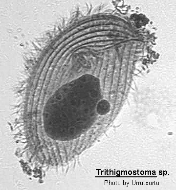
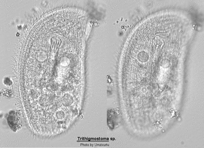
Outline shape oval to reniform, sometimes with and anterior left-hand rostrum. Dorso-ventrally flattened, ventral surface flat, dorsal surface arched except for an anterior flattened region which bears a row of bristles. Somatic ciliature mainly restricted to ventral surface, consisting of several longitudinal kineties on the right of which most curve around the anterior region of the body. The oral aperture is oval and is supported by a protrusible basket of trichites. There are 3 pre-oral kineties, and the middle one curves around the right side of the aperture. The pre-oral kinety furthest from the aperture lies along the suture line. The rest of the ventral surface is covered with longitudinal kineties and there is no central cilia-free zone as there is in Chilodonella. Macronucleus ovoid, many contractile vacuoles scattered throughout the cell.
Quote; Colin R. Curds "British and other freshwater ciliated protozoa Part I Ciliophora: Kinetofragminophora" Cambridge University Press, 1982 (ref. ID; 2013)
Body dorso-ventrally flattened; ventral surface flat with about 20 ciliary rows, the anterior part of the dorsal surface flattened with only 1 transverse row of cilia, the posterior part more less convex and lacking cilia; mouth opening round, about 12 cytopharyngeal trichites forming a tube, macronucleus oval with a characteristic concentric structure; 1 small micronucleus, about 6-8 contractile vacuoles. (ref. ID; 1219)
Measurements
Length 75-300 um, usually about 150 um. (ref. ID; 1219)
In vivo 70-100x30-40 um. Acontractile but highly flexible. Shape rather variable, similar to T. cucullulus, but more slender and right side often somewhat flattened at level of cytopharyngeal opening. Anterior end bluntly pointed and snout-like projecting, posterior end broadly to narrowly rounded. Dorsal hump sometimes projecting above flat ventral surface posteriorly. Praeoral area about 3:1, postoral portion about 2:1 flattened. Macronucleus in vivo about 25x13 um, in mid-body, contains spherical chromatin bodies surrounding hyaline area having central globule. Micronucleus conspicuous, in vivo about 7x5 um, adjacent to macronucleus. Usually 5-6 (rarely 8) contractile vacuoles along postoral body margins. Cytopharyngeal rods toothed, form dorsally directed funnel. Pellicle coated by an about 1 um thick, mucous layer. Cytoplasm colourless, with many 1-2 um sized, greasily shining globules and food vacuoles containing various diatoms. Movement slowly gliding, thigmotactic. Cilia of ventral side 5-6 um long. Somatic and oral infraciliature very similar to other members of genus. Distance between right and middle postoral kinety slightly enlarged, as in T. bavariensis. Kineties of right field successively shortened posteriorly. Dorsal brush subapical, near left margin, crosses snout, cilia 10 um long. (ref. ID; 4819)
Comparison with related species
The position of the dorsal kinety, the number and position of the contractile vacuoles, and the biotopes correspond with Sramek-Husek's findings. He obviously missed the slightly enlarged distance between the postoral kineties; it is in fact easily overlooked in vivo. Trithigmostoma srameki is not easily separated from some other members of the genus, especially from T. bavariensis. The only characters we found are the habitat (freshwater versus soil), the number of cytopharyngeal rods (11-14 versus 16-21) and the right ciliary field which is rounded posteriorly in T. srameki and transverse truncate in T. bavariensis (checked in three silver impregnated terrestrial populations). Trithigmostoma cucullulus (Muller, 1786) is slightly larger than T. srameki, has more (19-22) ventral kineties, and the distance between the postoral kineties is not enlarged. Furthermore, its dorsal brush consists of much more cilia (29-40, average 33). (ref. ID; 4819)
Neotype material
2 slides of protargol impregnated cells have been deposited in the collection of microscope slides of the Oberosterreichisches Landesmuseum in Linz, Austria. (ref. ID; 4819)