The World of Protozoa, Rotifera, Nematoda and Oligochaeta
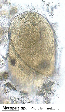
Metopus
Metopus Claparede & Lachmann, 1858 (ref. ID; 2014)
From Dr. InakiFrom Dr. InakiFrom Kuniyasu
Class Polyhymenophora: Subclass Spirotricha: Order Heterotrichida: Suborder Heterotrichina (ref. ID; 2014)
[ref. ID; 2014]
Body elongate, not flattened but asymmetrical due to the twisting of the anterior region to the left. Band-like AZM, situated in the anterior running obliquely around and down the body, terminating at the cytostome which is located either approximately equatorially of sub-equatorially. It never spirals down the body in an S-like manner. Without spines, armour or cirri. Jankowski considered there to be 2 subgenera depending upon the relative lengths of the anterior and posterior portions of the body. Sapropelic.
Most easily confused with Brachonella where the AZM is long, narrow and spirals down to the cytostome which lies in the posterior half of the body.
Quote; Colin R. Curds, Michael A. Gates and David McL. Roberts "British and other freshwater ciliated protozoa Part II Ciliophora: Oligohymenophora and Polyhymenophora" Cambridge University Press, 1983 (ref. ID; 2014)
[ref. ID; 4868]
The genus Metopus are characterized by torsion of the anterior part of the cell, and a frontal lobe which overhangs an obliquely ascending adoral zone of membranelles. The anterior twisting often gives Metopus a characteristic S-shape. All well-studied Metopus species have been shown to be anaerobic organisms, with a variable but always modest tolerance of dissolved oxygen. Although cells of some Metopus species may occasionally be found in oxygenated water, these are probably always accidental migrants. Representatives of the genus seem to be ubiquitous filter-feeders of bacteria in sediments, landfills and anoxic water. The history of the genus can be traced to the description of Trichoda es (syn. Metopus es) by O.F. Muller in 1786. The current generic name was erected by Claparede & Lachmann in 1858. Kahl (1935) discovered and named most of the currently known Metopus species. Thereafter, the genus continued to absorb new species, Corliss (1961) considered splitting it, and Jankowski (1964) carried out a partial taxonomic revision - allocating some species to the restored genera Bothrostoma and Cirranter, and others to the newly-created Brachonella and Tesnospira. Thus the remaining Metopus species became morphologically more homogeneous. The current status of the genus is that it contains 76 nominal species, and the obvious question is "why are these so many?". The following factors are probably relevant. Anaerobic ciliates are never very abundant and most species are relatively rare (at least in relation to aerobic ciliates). This means that observations of anaerobic ciliates in natural samples are often isolated sightings and it may be easier to give the different sightings different species names than it is to gain an impression of the spread in morphological variation of a natural population. Furthermore, until relatively recently, no anaerobic ciliates had been cultured, so the variation characteristic of anaerobic ciliate species was rarely known. When we include some other relevant factors: (a) the existence of polymorphic anaerobic ciliates (the most spectacular example being M. palaeformis), (b) species nomenclature based on differences in gross cell morphology rather than the more conservative somatic and oral ciliature, (c) the physical fragility of many anaerobic species and the ease with which aberrant specimens can be produced (and described), and (d) some obvious cases of ignorance of previously-published literature, it becomes clear that too many species have been erected for the variety of Metopus that have been observed so far. All Metopus species fall with five broad categories of cell shape. Kahl (1935) too divided Metopus into five groups of species, according to general cell morphology and the size of the AZM (his Group VI is currently represented by the genus Brachonella). The great morphological variety which characterizes many Metopus species, allows them to be placed in groups based solely on cell shape. The groups are as follows:
Group I: Metopus palaeformis-like organisms. The equatorial part of the cell is as wide as or narrower than the posterior part. These ciliate are usually thin, and elongate.
Group II: Metopus striatus-like organisms. Bell-shaped organisms, resembling spine-less Caenomorpha. Cells are wider at the anterior and equatorial parts of the cell than in the posterior half. With or without caudal protuberance or projection; when present, this projection is not spine-like, but a round short projection.
Group III: Cells are wider at the equator than at the anterior and posterior ends. The posterior end can vary between species, from very narrow (e.g. M. es) to less so (e.g. M. contortus).
Group IV: Approximately ovoid cells (e.g. M. ovalis).
Group V: Metopus with the posterior part of the cell narrower than both the cell equator and the anterior end, and with a conspicuous and distinctive spine-like posterior extension, e.g. M. vestitus.
The genus Metopus are characterized by a twisted anterior part that includes 5 to 10 kineties, some of them closer to each other at the edge of an overhanging lobe, where they form the so-called perizonal ciliary stripe (Jankowski 1964). The perizonal are runs above the course of the adoral zone of membranelles (AZM), which reaches the equator of the cell in most species. The overhanging lobe is not always prominent, but its presence together with the torsion of the cell, makes most of the ciliates in the genus S-shaped. Most Metopus species have a group of distinctive intracellular dark particles close to the anterior end of the cell. The size and shape of some Metopus species vary greatly with changes in physiological state. (ref. ID; 4868)
In the digestive tract of sea urchins, Diadema setosum and Echinometris subangularis in Bermuda; in Centrechinus antillarum, etc. in Tortugas; in Diadema setosum and Echinometra oblonga in Japan. (ref. ID; 1618)
M. contortus has the typical Metopus shape: wide at the cell equator, with a narrow peristome in the anterior part. The living organism is 89-165 um long, and 26-51 um wide. The somatic infraciliature is constituted by about 40 kineties. The kinetosomes in the kineties are paired, and each bears a cilium. The peristome has five kineties, of which two or three are closer to each other. The AZM is long, with 35-50 membranelles. The cytostome opens behind the cell equator. The macronucleus is single and situated in the anterior half of the cell. The micronucleus is placed in a depression of the macronucleus. The contractile vacuole is single and close to the posterior end of the cell. Caudal cilia are present. The cytoplasm contains endosymbiotic methanogenic bacteria. The external surface bears ectosymbiotic bacteria which are probably sulphate reducers (Fenchel & Ramsing 1992). M. contortus is easily isolated in anaerobic culture from marine sediments. It is common in almost all types of anaerobic marine habitats, including the anoxic water column, sediments, bacterial mats and decaying sea weeds. (ref. ID; 4868)
Body form oblong sigmoid; the slightly spiral diagonal peristome divides the body into anterior and posterior portions; body ciliation uniform; the longitudinal rows of cilia in the posterior part of the body curve into the anterior part on account of the sigmoid torsion; the peristome is surrounded anteriorly by the perizonal ciliary stripe, which includes 5 ciliary rows and is equipped with about 32 membranelles; a small undulating membrane on the right upper side of the cytostome; 1 oval macronucleus and 1 micronucleus near the peristome; 1 terminal contractile vacuole; refractile bodies accumulated near the anterior pole. (ref. ID; 1219)
It is a very distinctive S-shaped ciliate. 112-203 um long, and 23-66 um wide, with a protruding anterior part and a characteristically elegant motion when swimming. There are about 35-40 somatic kineties, of which 5 form the perizonal stripe. The AZM includes about 40-50 membranelles. The endosymbiotic methanogens are found throughout the cytoplasm. The single macronucleus is more or less ovoid and placed in the anterior part, close to the equator of the cell. The single micronucleus lies next to the macronucleus. There are two short caudal cilia which are only observable on stationary living organisms. This is a very common ciliate in anoxic freshwater habitats. (ref. ID; 4868)
Metopus halophila is 50-90 um long, ellipsoidal with the anterior part of the cell projecting forwards, when the cell is seen from the side. This feature confers a curved appearance, closer to an open C-shaped than the typical Metopus S-shape. Small ectobiotic bacteria cover most of the external cell surface. The ciliates have about 20 somatic kineties with paired kinetosomes and every kinetosome bears a cilium. There is also a group of four caudal cilia. The contractile vacuole is terminal and very obvious. The oral region is shorter than in other Metopus species, and does not reach the equator of the cell. This oral region is curved towards the dorsal side, following the shape of the protruding anterior part. The AZM includes 12-15 membranelles. Metopus halophila is not very common and, along with M. vestitus, occurs in the anaerobic layer of marine sand and in bacterial mats. (ref. ID; 4868)
Size in vivo usually 80-130x15-30 um. Overall shape cylindroidal; preoral dome slender and indistinctly projecting above ventral surface, inclined about 45 degrees to main body axis projecting distinctly above left body margin. Macronucleus ellipsoidal and usually in anterior body half left of adoral zone of membranells. Cortical granules inconspicuous. Five perizonal and usually 10-15 somatic ciliary rows, of which two to three extend onto preoral dome. About five distinctly elongated caudal cilia. Adoral zone occupied circa 35-45% of body length on average, terminates on ventral side right of midline slightly underneath perizonal stripe, composed of about 16-24 membranelles. (ref. ID; 2884)
Descriptions
Overall shape cylindroidal, length:width ratio highly variable within and between populations both in vivo and protargol slides, where M. hasei is considerably stouter than when alive: in vivo 2.5-6:1, usually 4:1-5:1; after protargol impregnation 1.8:1-7.6:1, usually 2.7:1-4.9:1. Ratio of preoral:postoral body portion also rather variable, usually about 1:1-1:1.9. Preoral dome only slightly sigmoidal, distinctly curved and inclined about 45 degrees to main body axis, projecting knob-like above anterior left body margin; inconspicuous because without distinct brim, narrow, dorsoventrally flattened about 2:1, and hardly projecting above ventral surface merging smoothly into right side in middle third of cell. Postoral body portion cylindiroidal with rear end slightly narrowed and evenly rounded, frequently with more or less distinct folds, especially after systole of contractile vacuole. Macronucleus typically in anterior body half left of adoral zone of membranelles, in vivo about 28-36x8-14 um, that is, short to long-ellipsoidal, ovoidal or reniform, length:width ratio usually about 2-4.5:1; contains numerous nucleoli 0.6-1.5 um across. Micronucleus usually near or attached to anterior third of macronucleus, globular to ellipsoidal, surrounded by distinct membrane in alpine population. Contractile vacuole in posterior end, with very short canal extending to argyophilic cytopyge slit on posterior pole, indicating that a separate excretory pore is lacking, as in M. inversus. Cortex flexible, slightly furrowed by ciliary rows, contains colourless granules difficult to recognize in vivo but occasionally distinct in protargol and methyl greenpyronin preparation, being rather densely arranged and 0.2-0.7 um across; tightly underneath cortex pale, ellipsoidal granules, possibly hydrogenosomes. Cytoplasm colourless, hyaline, especially in posterior body portion, bacterial rods, probably methanogens, impregnate with protargol and/or silver carbonate in some populations. Food vacuoles 4-14 um across, contain bacteria. Movement moderately fast by rotation about main body axis. Normal somatic cilia 10 um long, those of perizonal stripe and underneath buccal vertex elongated to 13 um. Invariably about four to six caudal cilia, whose length varies considerably in different populations: as long as body, about 30 um, 35 um in live specimens from a riparian forest soil in Lower Austria, 30-45 um (average=38.8 um, n=10) in protargol-impregnated cells from Namibia, and 30-76 um (average=50 um, n=19) in protargol-impregnated specimens for South Africa. Number of ciliary rows rather variable, about 15 in type population and in Namibian and South African specimens, only 10 in the alpine population from Austria. Ciliary rows slightly shortened posteriorly, leaving black a small, roughly circular area containing the cytopage, every second to third row elongated by one dikinetid bearing a caudal cilium; composed of dikinetids orientated parallel to slightly obliquely to kinety axis and having only the posterior basal body ciliate, except the anterior portion of the dome and postcytostomial kineties and the whole perizonal ciliary rows, where both basal bodies are ciliferous; longitudinally and equidistantly arranged in Namibian and South African specimens, slightly narrower spaced dorsally than ventrally in Austrian population, where often a rather distinct glabrous stripe occurs in midline. Postoral (ventral) kineties distinctly separate from adoral zone of membranelles, dorsal kineties anteriorly shortened from right to left. Two dome kineties in Austrian, three in Namibian and South African specimens, each composed of about 18-30 dikinetids. Perizonal stripe slightly shorter than adoral zone of membranelles, about 2-3 um wide, composed of five narrowly spaced kineties following curvature of dome margin; kineties 4 and 5 slightly shortened anteriorly and posteriorly and slightly apart from and more widely spaced than kineties 1-3. Perizonal dikinetids widely spaced, those of rows 1-3 from 20-36 short, straight ("false") kineties alternately arranged to dikinetids of kineties 4 and 5, producing arrow-like pattern. Somatic dikinetids associated with four conspicuous structures after silver carbonate impregnation: anterior basal body associated with structure (a) directed to the left and slightly anteriad; posterior basal body associated with structure (c) extending obliquely posteriad, an anterolaterally-directed structure (d) on the right, an a structure (b) extending to the left and very slightly posteriad, forming an acute angle with structure (a) of the anterior basal body. The length of the associates depends on the body region and is especially diverse in the perizonal ciliary stripe, producing its peculiar texture after silver carbonate impregnation. Structures (a, b) are distinctly longer than structure (c), both are very short in the perizonal stripe, except for structure (c) in row 1, which extends between the "false" kineties. Structure (d) is about as long as structure (c) in the postoral body portion, its length distinctly increases anteriad in the dome kineties and, especially, in the mid-ventral portion of perizonal ciliary row 5. Adoral zone of membranelles slightly sigmoidal, hardly roofed by preoral dome, commences at anterior left margin of preoral dome and extends obliquely to right body margin, performing a slight clockwise rotation when plunging into the shallow buccal cavity slightly above mid-body; consists of 17-20 membranelles in Austrian, 15-18 in Namibian, and 21-26 in South African population. Zone composed of a long distal and a very short proximal portion distinctly different in the structure of the membranelles: distal membranelles cuneiform because composed of two long (2-3 um) rows of zigzagging basal bodies to which a short row is attached at right anterior end; proximal (buccal) membranelles rectangular and composed on only two rows of basal bodies. Paroral membrane in corner formed by preoral dome and ventral surface, short and almost straight, extends to proximal end of buccal cavity, composed of basal bodies in single line bearing about 10 um long cilia, appears rather thick in silver slides due to adhering (pharyngeal) fibres, which form dorsally directed funnel extending to near posterior body end. (ref. ID; 2884)
Comments
Metopus hasei is similar to M. palaeformis Kahl, 1927 in shape, size, and number of ciliary rows (8-14) and adoral membranelles (10-20). Unfortunately, Esteban et al. did not provide details of the infraciliature of M. palaeformis. In spite of this, M. hasei and M. palaeformis can be easily separated because the latter lacks caudal cilia. The South African specimens of M. hasei are very similar to M. laminarius f. minor Kahl, 1932, indicating synonymy. However, a reliable comparison is impossible because the infraciliature of both M. laminarius (length 200-250 um) and M. laminarius f. minor (150 um) is not known. (ref. ID; 2884)
Size in vivo about 80x50 um. Mushroom-shaped due to laterally and ventrally widely projecting preoral dome and narrowed, ellipsoidal postoral body portion. Dome conspicuously sigmoidal and flattened, contains reniform macronucleus. Cortical granules inconspicuous. Five perizonal and 24 somatic ciliary rows on average, of which 4 extend onto preoral dome. Adoral zone slightly shorter than perizonal stripe, terminates in mid-body on right margin of cell, composed of 33 adoral membranelles on average. (ref. ID; 2884)
Descriptions
Size in vivo 70-100x40-70 um, usually about 80x50 um, as calculated from some measurements of live specimens; length:width ratio highly variable (1.2-2.3:1), usually 1.7:1, ratio of preoral:postoral body portion also rather variable, on average about 1:1. Overall shape mushroom-like, it depends, however, on the side viewed due to the widely over-hanging and sigmoidally ascending preoral dome occupying about one fifth of body length in ventral view. Preoral dome, although thin and hyaline, very conspicuous because widely projecting above ventral and lateral body surface, extends almost perpendicularly above ventral side and merges into body proper slightly underneath mid-body on right margin of dorsal side; central dome portion slightly convex, broadly sigmoidal in top view due to curved dome brim, which forms sharp corner with dorsal side anteriorly. Postoral body portion (mushroom stem) ellipsoidal with ventral and left side more distinctly convex than right and dorsal, causing a slight, sigmoidal torsion of the organisms, described rather cryptically by Jankowski as "sigmoid body with double winding". Rear third of body with some more or less pronounced folds after systole of contractile vacuole. Macronucleus invariably in preoral dome, reniform, contains numerous nucleoli. Micronucleus ellipsoidal, usually near or attached to ventral anterior end of macronuleus. Cytopyge subterminal on ventral side, slit-like, very likely also functioning as discharge device for the contractile vacuole because no excretory pore could be found. Cortex slightly furrowed by ciliary rows, contains inconspicuous, loosely arranged, colourless granules 0.2-0.5 um across, which stain red with methyl green-pyronin. Cytoplasmic bacteria neither recognizable in vivo nor after silver carbonate and protargol impregnation; no particular accumulation of granules in preoral dome. Food vacuoles 5-15 um across, contained bacteria and their spores. Movement moderately fast, without peculiarities. Somatic cilia about 15 um long in vivo, lack of elongated caudal cilia checked in live and over-impregnated specimens. Ciliary rows composed of dikinetids oriented parallel to slightly obliquely to kinety axis and having only the posterior basal body ciliated, except perizonal ciliary rows and anterior portion of dome kineties, where both basal bodies are ciliferous; longitudinally and equidistantly arranged underneath membranellar zone, dorsally distances increase from right to left. Postoral (ventral) kineties distinctly separate from adoral zone of membranelles, slightly shortened posteriorly, leaving black a small, roughly circular area containing the cytopyge. Dorsal kineties anteriorly shortened from right to left, while slightly elongated posteriorly and thus extending across pole to circular patch containing the cytopyge. Accordingly, the posterior cell pole does not entirely coincide with the kinety pole, which is subterminally on the ventral side. Ciliary pattern rather irregular underneath buccal vertex and at posterior end of perizonal stripe, where some scattered dikinetids occur. Three to four dome kineties, number 1 as long as perizonal stripe, numbers 2-4 slightly shortened anteriorly and extending to posterior body end; distances between dome kineties decrease from ventral to dorsal. Perizonal ciliary stripe slightly longer than adoral zone of membranelles, about 3 um wide, composed of five very narrowly spaced kineties following curvature of dome margin, kineties 4 and 5 slightly shortened anteriorly and posteriorly and slightly apart from and more widely spaced than kineties 1-3. Perizontal dikinetids widely spaced, those of rows 1-3 form 58-74 short, straight ("false") kineties alternately arranged to dikinetids of kineties 4 and 5, producing arrow-like pattern. The associates of the somatic dikinetids and their orientation match those of M. hasei. Likewise, the length of the associates depends on the body region and is especially diverse in the kinetids of the perizonal stripe, producing its peculiar texture after silver carbonate impregnation. Structures are longest in the posterior half of the postcytostomial ciliary rows and in the anterior half of the dorsal and dome kineties, except the anteriormost one to two dikinetids. Structure (d) is elongated in the anterior dome area, increasing in length from posterior to anterior and from dorsal to ventral and, especially, in the mid-ventral portion of perizonal ciliary row 5. In the perizonal ciliary rows most associates are short, except structure (d) of row 5; structure (a) of rows 2 and 5; structures (a, b) of row 4, which extend to row 3 and diverge so strongly that each touches one of the short, "false" kineties; and structure (c) of row 1, which extends between the short "false" kineties. Adoral zone of membranelles sigmoidal, entirely roofed by dome brim, commences at anterior dorsal end of preoral dome and extends obliquely to mid-body and right side, where it plunges into the short buccal cavity performing a slight clockwise rotation. Zone composed of along distal and a short proximal portion which differ distinctly in the structure of the membranelles: distal membranelles cuneiform because composed of two, about 3 um long rows of zigzagging basal bodies to which a short row is
attached at right anterior end; proximal (buccal) membranelles rectangular and composed of four rows of basal bodies, those in mid of buccal cavity slightly longer (about 5 um) than all other membranelles. Paroral membrane in corner formed by preoral dome and ventral surface, commences near midline of cell and curves to proximal end of buccal cavity, composed of ciliated basal bodies in single line, appears rather thick in silver slides due to adhering (pharyngeal) fibres, which form long, dorsally directed funnel with fibres widely spaced in aperture region. (ref. ID; 2884)
Comparison with original description and related species
Metopids have a complicated shape and thus many different aspects. We identified our population as Brachonella inversa because the two (uncommon) views Jankowski showed almost perfectly march some of our illustrations, and both populations have the macronucleus in the preoral dome and lack caudal cilia. Furthermore, the size of preserved specimens (63-91x38-61 um vs. 90-105x51 um) and the long perizonal stripe agree with Jankowski's description. However, there are (minor) differences, namely the perizonal stripe, which is slightly shorter than the adoral zone of membranelles in our population, and the dome brim, whose hook-like dorso-lateral projection was not described by Jankowski. Both characters are difficult to recognize without video microscopy and silver impregnation. Thus, it is reasonable to assume that he overlooked them. In vivo, B. inversa looks like a stoutish Metopus hasei. However, both are easily distinguished by the location of the macronucleus (in preoral dome vs. postoral body portion) and the caudal cilia (lacking vs. present). In silver preparations, they differ by the number of somatic kineties (22-25 vs. 10-16). The identification of metopids is difficult, mainly because very few species have been thoroughly studied and no type material is available. (ref. ID; 2884)
Generic classification
Jankowski split Metopus into several genera and subgenera. Brachonella differs from Metopus mainly by the subterminal, dorsal location of the cytostome due to the strongly spiralized adoral zone of membranelles. Jankowski's figures of B. inversa largely agree with our observations and show that the adoral zone of membranelles hardly extends onto the dorsal side and certainly not to the posterior body end. This contradicts his description, at least partially, which is based on unstained, mercuric chloride preserved specimens: "The elongated adoral zone of membranelles makes a spiralling band, that shifts the cytostome on the dorsal body surface to the posterior end. This spiral is much longer in B. spiralis; in this respect, B. inversa occupies an intermediate position between Metopus es and Brachonella spiralis". According to our investigation, B. inversa is a typical member of Metopus because the cytostome is near mid-body (Jankowski did not recognize that the adoral zone is slightly shorter than the perizonal stripe) and in the transition zone of the right and dorsal side. Accordingly, we combine Brachonella inversa Jankowski, 1964 to Metopus inversus nov. comb. Note that Jankowski added "sp. n." to M. inversus in two publications, which appeared in the same year. However, only in one of the two studies he provided also a description; thus, this paper should be considered as the place of the original description of M. inversus. (ref. ID; 2884)
M. major is larger: 113-191 um long and 26-49 um wide. The somatic infraciliature includes 50 to 55 kineties. The AZM consists of 80 membranelles, and in some specimens it extends far beyond the cell equator. The perizonal area is formed by at least 5 or 6 kineties lying very close to each other. The contractile vacuole is close to the posterior end of the cell and a group of caudal cilia is also present. In living organisms these caudal cilia are always straight and stiff. The macronucleus is elongate and placed in the centre of the cell. The micronucleus does not lie next to the macronucleus, but in the posterior half of the cell. (ref. ID; 4868)
Type locality
M. major was isolated from a sulphidic accumulation of decaying eelgrass in Niva Bay (Denmark). (ref. ID; 4868)
The ciliate is elongate, ellipsoid, and 80-100 um long, with a broad perizonal area. The AZM extends from the left anterior part of the cell, twisting rightwards in an S-form to beyond the cell equator. It includes 32-35 membranelles. The anterior part of the cell is wide, compared to other Metopus species. The somatic infraciliature is formed by 50 kineties, all with paired kinetosomes bearing cilia. Protargol-stained organisms also show at least four kineties closer to each other in the anterior part of the cell, forming the perizonal ciliary stripe. M. nivaaensis has seven to 10 caudal cilia in a group. There is a single contractile vacuole placed close to the posterior end. The single macronucleus is in the centre of the cell, and the micronucleus is placed above it. M. nivaaensis is common in anaerobic marine sands and in masses of purple sulphur bacteria. The ellipsoidal shape, and the wide anterior part make M. nivaaensis easily distinguishable from other Metopus species. (ref. ID; 4868)
Type locality
It was isolated from Niva Bay (Denmark). (ref. ID; 4868)
This ciliate is polymorphic and it is probably the most variable of the Metopus spp. in culture. A typical trophic cell is elongate, flattened, and somewhat ribbon-like in shape. Cell size varies from 70 to 200 um long (exceptionally, longer), and 8-31 um wide. The number of somatic kineties is 8 to 14, and there are 10-20 membranelles in the adoral zone. The somatic kineties are slightly curved in the posterior part of the cell and they are formed by dikinetids which have the same ultrastructure as the dikinetids in M. contortus. The arrangement of somatic kineties in smaller specimens of M. palaeformis resembles that in some species of Trachelophyllum. All forms of M. palaeformis (including the cysts) have endosymbiotic methanogenic bacteria distributed throughout the cytoplasm. These endosymbionts do not undergo morphological transformation. They are rod-shaped, and they lie close to the hydrogenosomes (Finlay & Fenchel 1991). Mucocysts lie beneath the entire cell surface, including the oral region. M. palaeformis is a common freshwater ciliate. It has also been found in samples of anaerobic municipal landfill material (Finlay & Fenchel 1991), in a septic tank, in sulphide-rich solution lakes, in soil from an extinct volcano, and in marine lagoons (Aladro et al. 1990). Due to the wide morphological variation now known for M. palaeformis, we conclude that the species is probably the same thing as the Tesnospira described by Jankowski (1964), as well as Metopus hyalinus Kahl, 1927, M. rostratus Kahl, 1927, and M. tenuis Kahl, 1927. (ref. ID; 4868)
These ciliates are characterized by curvature of the cell, which is especially conspicuous in the living organism, and the presence of a caudal prolongation or spine. The latter varies in length and in some individuals it is very short. The curved shape, together with the caudal spine confers a peculiar swimming movement: the posterior half of the cell moves alternately from one side to the other. The ciliates are approximately 70 um long, and always wider at the cell equator. Our observations agree with those of Jankowski (1964) for M. caudatus Da Cunha, 1915. The contractile vacuole is situated at the cell posterior and is cuboid to ellipsoid. In agreement with Jankowski (1964), we have observed long (6-9 um) intranuclear rod-shaped bacteria, of unknown identity. The cytoplasm of M. spinosus is very transparent, usually containing groups of pink spherical particles -possibly ingested purple bacteria. The cytoplasm bears other rod-shaped bacteria, which we also failed to identify. They did not show typical methanogen autofluorescence. The macronucleus is spherical and situated in the centre of the cell. The AZM includes about 15 membranelles; the somatic infraciliature is formed by less than 20 kineties, each with a few sparsely-located cilia, and two cilia per dikinetid. There are no caudal cilia. We have encountered M. spinosus on several occasions in the anaerobic sediment of freshwater ponds in the U.K. The caudal prolongation or spine is variable in morphology. We have observed organisms with a completely developed caudal prolongation; other specimens had a little protuberance at the base of the caudal spine, as described by Kahl (1935) for M. spinosus, and by Jankowski (1964) for M. caudatus. There are also spine-less organisms that look like M. curvatus and M. convexus described by Kahl (1935). Taking account of all these observations we have decided to name this ciliate M. spinosus Kahl, 1927, although the same thing was described by Jankowski (1964) as M. caudatus Da Cunha (1915). Da Cunha found the organisms he described in marine habitats and they probably correspond to M. vestitus (Kahl, 1927). Accordingly, M. vestitus should be renamed M. caudatus, as the latter was first described. To avoid more confusion we leave M. vestitus as it is (Kahl's description is very precise), with M. caudauts as a synonym, and we call our freshwater Metopus, M. spinosus, with synonyms M. curvatus, M. convexus, and M. attenuatus. (ref. ID; 4868)
This distinctly tear-shaped ciliate is 38 to 71 um long, with a pointed posterior end bearing caudal cilia. The number of kineties is about 25, although it may vary with cell size. Five somatic kineties lie closer to each other in perizonal region. The AZM is formed by about 45 membranelles. The ciliates bear endosymbiotic methanogenic bacteria. The macronucleus is single, rounded, and located in the centre of the cell, the micronucleus lying close to it. Extrusomes lie beneath the cell membrane, although they are not always obvious. When the ciliate is found in nature it has a well-developed caudal protuberance, as explained above. After several weeks in culture, the cells become smaller and more rounded, and they lose the caudal projection. The shape of the macronucleus remains constant. M. striatus is quite common in soft sediments in freshwater ponds and lakes as well as in river sediments. (ref. ID; 4868)
Measurements
80-120 um long. (ref. ID; 1618)
Metopus verrucosus Da Cunha, 1915 (ref. ID; 1620, 1621) or (Da Cunha, 1915) Kahl, 1935 (ref. ID; 4868)
Descriptions
This ciliate is rare and uncommon but it can be found in Beggiatoa mats in marine habitats. The ciliate fits the descriptions of Da Cunha (1915) and Kirby (1934). Kahl (1935) included this organism (Spirorhynchus verrucosus) within the genus Metopus. It is 100-140 um long and about 20 um wide, elongate and thin, with both ends tapered, long cilia, and a posterior contractile vacuole. It seems to have three macronuclei and no micronuclei (Kirby 1934), although variations in this number might be possible (Da Cunha 1915 did not observe nuclei). The principal feature of this organism are the tufts of ectobiotic bacteria. These are grouped over the surface of the ciliate except in the narrow anterior region. We have encountered it a few times but have failed to photograph it. In recent years is has become clear that truly anaerobic free-living ciliates do exist, that the majority of them live in symbiotic association with methanogens and/or sulphate reducers, and that together with other free-living anaerobic protozoa, they are the only important predators in the typically short food chains of anaerobic environments. The natural history of some of these ciliate cosortia is described in recent reviews (Finlay & Fenchel 1993; Fenchel & Finlay 1995). While much effort has recently been devoted to describing aspects of the genotype (especially small sub-unit ribosomal rRNA sequences) of some anaerobic ciliates and their symbionts (Embley & Finlay 1994), and to describing new species with novel types of symbiotic consortia (e.g. Esteban et al. 1993), very little attention has been directed towards the diversity of anaerobic ciliates, as a whole. The taxonomy of these organisms has its foundations in the seminal works of Kahl (1935) and Jankowski (1964), but in the last 10 years or so it has become possible to culture some of these organisms, and we now know a great deal about them. Now we also know something about the distribution and abundance of these organisms in the natural environment (Finlay et al. 1991; Fenchel & Finlay 1995; Fenchel et al. 1995), and the time is ripe to take a critical look at the diversity of anaerobic ciliates. The current offering has dealt solely with the genus Metopus. (ref. ID; 4868)
The ciliate is S-shaped, about 70 um long, with a distinctive, long, spine-like posterior extension. The ciliates bear a coat of ectobiotic bacteria. These divide transversally. There are about 25 somatic kineties, with paired kinetosomes. The oral region consists of 20 oral membranelles. The macronucleus is single and elongate, with one or two associated micronuclei. The ciliates bear endosymbiotic methanogens. In the anterior part of the cell and in the posterior projection there are needle-shaped intracytoplasmic structures, which were observed by Tucolesco (1962). These appear to the crystals, and they resemble structures described in other marine ciliates (Dragesco 1960). M. vestitus is not very common. It occurs in anaerobic layers of marine sand, and sporadically in other marine benthic habitats such as bacterial mats. (ref. ID; 4868)