Lembadion
Lembadion Perty, 1849 (ref. ID; 2014) or 1852 (ref. ID; 3690)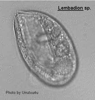
Hymenostomata (ref. ID; 4831) [ref. ID; 2014]
More or less ovoid ciliate, ventral surface flat to slightly concave, dorsal surface convex. Very large oral aperture occupying about three-quarters of the length of the ventral surface beginning at the apical pole with the cytostome at the posterior end. There is a long undulating membrane on the right which is insignificant when compared with the very large membranelles on the left. These membranelles which dominates the ventral such as to appear as a single large membranelle which dominates the ventral surface. Somatic ciliation uniform arising from a clearly sculptured pellicle and these is usually a tuft of caudal cilia. The contractile vacuole is dorsally located and is connected to the excretion pore via a long curved canal. The macronucleus is an elongate ovoid lying in a posterior position with a single spherical micronucleus.
Quote; Colin R. Curds, Michael A. Gates and David McL. Roberts "British and other freshwater ciliated protozoa Part II Ciliophora: Oligohymenophora and Polyhymenophora" Cambridge University Press, 1983 (ref. ID; 2014)
- Lembadion bullinum (Mueller, 1786) Perty, 1849 (ref. ID; 4488, 4611) or Perty, 1849 (ref. ID; 4831), 1852 (ref. ID; 1308, 1335, 1622, 1629, 2245, 4305) reported year? (ref. ID; 1618, 3698) reported author and year? (ref. ID; 191)
Syn; Bursaria bullina O.F. Muller, 1786 (ref. ID; 4611) - Lembadion bullinum Perty var. arenicola Dragesco, 1960 (ref. ID; 3690 original paper)
- Lembadion conchoides Faure-Fremiet, 1924 (ref. ID; 1622) reported author and year? (ref. ID; 4611)
See; Lembadion magnum (ref. ID; 1622) - Lembadion duriusculum
See; Cristigera phoenix Penard, 1922 (ref. ID; 1622) - Lembadion lucens (Maskell, 1887) (ref. ID; 1622, 1629, 1896, 4305) reported year? (ref. ID; 3698, 5462) or (Maskell, 1887) Kahl, 1931 (ref. ID; 4488, 4611) reported author and year? (ref. ID; 191, 3954)
Syn; Thurophora lucens Maskell, 1887 (ref. ID; 4611) - Lembadion magnum (Stokes, 1887) (ref. ID; 1219, 1622, 1629) reported year? (ref. ID; 5462) or (Stokes, 1887) Kahl, 1931 (ref. ID; 4611) reported author and year? (ref. ID; 191, 7768)
Syn; Hymenostoma magna Stokes, 1887 (ref. ID; 4611); Lembadion conchoides Faure-Fremiet, 1924 (ref. ID; 1622) - Lembadion magnus (ref. ID; 3954)
- Lembadion ovale Gourret & Roeser, 1886
See; Lembus pusillus (ref. ID; 1622)
Lembadion bullinum (Mueller, 1786) Perty, 1849 (ref. ID; 4488, 4611) or Perty, 1849 (ref. ID; 4831), 1852 (ref. ID; 1308, 1335, 1622, 1629, 2245, 4305) reported year? (ref. ID; 1618, 3698) reported author and year? (ref. ID; 191)
Synonym Bursaria bullina O.F. Muller, 1786 (ref. ID; 4611)Descriptions
See the description of Lembadion lucens. [ref. ID; 4831]Measurements
120-200 um long; posterior cilia 40-50 um long. (ref. ID; 1618)A comparison of the biometrical data of normal cells and giants clearly shows the differences in cell size: Whereas the cell length of normal cells used for the study varied from 96 to 136 um, well fed giants from the same culture had a length of 142-199 um and still 132-190 um if slightly starved. This means that giants, in a stage when they feed on normal cells, are completely separated from them in cell length and that these giants only lose less than 10% of their length during three days of starvation (but more than 25% of their width). In conclusion, the cell length proved to be a suitable criterion for the definition of a giant in comparison to normal individuals present in the same culture. The size of the buccal cavity also has increased in giants. Whereas its average length is about 90 um in normal cells, average lengths of 140 and 130 um have been measured for well fed and slightly starved giants, respectively. This means that a normal cell (in the average 116 um in cell length) can easily be ingested by a giant, even if the prey is turned on its long axis inside the buccal cavity of its predator. (ref. ID; 4831)
Lembadion lucens (Maskell, 1887) (ref. ID; 1622, 1629, 1896, 4305) reported year? (ref. ID; 3698, 5462) or (Maskell, 1887) Kahl, 1931 (ref. ID; 4488, 4611) reported author and year? (ref. ID; 191, 3954)
Synonym
Thurophora lucens Maskell, 1887 (ref. ID; 4611)Descriptions
- General morphology: Lembadion lucens and L. bullinum are oval-shaped freshwater ciliates. Both species have, at their posterior poles, a tuft of long caudal cilia characteristic of this genus. The nuclear apparatus, located in the posterior part of the cell, consists of an elongated macronucleus and one spherical micronucleus close to it. However, L. bullinum is bigger than L. lucens. (ref. ID; 4305)
- Somatic infraciliature: The somatic infraciliature of L. lucens is made up of 30-35 meridional kineties, while L. bullinum possesses 50-60 somatic kineties. In both species, the majority of the somatic kineties are located on the dorsal side of the ciliate resulting in the extension of the buccal cavity. Moreover, left and right ventral kineties converge to form a postoral suture behind the mouth, which is bigger in L. bullinum. At light microscopical level, the somatic kineties of the two species seem to be composed of dikinetids, each one with a long kinetodesmal fiber extending toward the next anterior kinetosome. The infraciliature of the caudal cilia in L. lucens and L. bullinum consists of two monokinetid rows: one of them is made up of the last kinetosomes of the 1st ventral kineties, and the other row is formed by the last kinetosomes of the 1st dorsal left kineties. In L. lucens, the two rows are composed respectively of six and four kinetosomes separated by two or three somatic kineties, while in L. bullinum these rows has respectively 10-15 and five caudal kinetosomes separated by three or five somatic kineties. The argyrome, only stained in L. lucens, appears as a quadrangular network in which the kinetosomes are slightly displaced to the right. (ref. ID; 4305)
- Buccal infraciliature: The left margin of the buccal cavity is limited by one polykinety (Pk), and the right margin is surround by two paroral kineties (PK1 and PK2). The polykinety is made up of seven files of kinetosomes in L. lucens, while it consists of nine kinetosomal files in L. bullinum. The most external files of the polykinety, three in L. lucens and five in L. bullinum, begin and finish at different levels. In both species, the paroral kineties begin at the same level in the apical pole of the cell, but only the outer paroral kinety (PK1) reaches the posterior end of the mouth. At light microscopic level, the infraciliature of the two paroral kineties seems to be different: the outer paroral kinety (PK1) seems to be composed of pairs of kinetosomes, and the inner one (PK2) of isolated kinetosomes; however, electron microscopy shows that both paroral kineties are formed by kinetosome pairs (Bardele, pers. commun.). The bottom of the buccal cavity of L. lucens and L. bullinum shows three longitudinal argentophilic structures: a strongly argentophilic fiber (L1) located at the right side; a granular line (L2) situated at the left side and separated about 2 um from he polykinety; and a thin line showing a zigzag pattern (L3) located between the other two aforementioned lines. Moreover, the buccal cavity is crossed by thin transverse striations that can be differentiated in three zones: one zone (St1) occupies the major part of the buccal cavity, and their striations begin in the granular line (L2) and finish at 3 um of the zigzag line (L3); another zone (St2) is situated between the zigzag line (L3) and the argentophilic fiber (L1); and the last one (St3) is observed between the argentophilic fiber (L1) and paroral kineties (PK1 and PK2). (ref. ID; 4305)
- Binary fission. Stomatogenesis: The stomatogenic process of L. lucens and L. bullinum can be subdivided in five stages:
- Stage 1. First sign of stomatogenesis is the proliferation of the inner paroral kinety (PK2) at its posterior zone, giving rise to one small file of kinetosomes located between the two paroral kineties (PK1 and PK2).
- Stage 2. The PK2 proliferation continues towards the anterior part forming the primordium of the new polykinety (Pk') constituted by one file of kinetosomes. The proliferation of the outer paroral kinety (PK1) begins at this stage.
- Stage 3. The primordium of the new polykinety (Pk') proliferates by elineation, reaching the characteristic number of files for each species, seven in L. lucens and nine in L. bullinum. The proliferation of the outer paroral kinety (PK1) continues according to a postero-anterior gradient.
- Stage 4. The proliferation of the outer paroral kinety (PK1) originates in two files of kinetosomes which will form the new paroral structures of the opisthe (PK'1 and PK'2). Simultaneously, a 2nd proliferation of the inner paroral kinety of the parental cell (PK2) takes place regenerating the outer paroral kinety of the proter (PK1).
- Stage 5. Adoral and paroral structures of the opisthe, located at right side of the old mouth, displace towards the posterior pole and separate from one another. At the same time, the longitudinal fiber (L1) separates from the inner paroral kinety in both daughter cells.
- Binary fission. Somatic infraciliature: The proliferation of the somatic infraciliature starts slightly before the stomatogenic process at a subequatorial level. This proliferation leads to groups of 3, 4, 6 or more packed kinetosomes which form a ring surrounding the cell. Simultaneously with stomatogenesis, the somatic proliferation extends anteriorly and posteriorly forming a proliferation band in the dividing cell. The basal bodies then separate. In the later stages of the fission process, when the scission furrow is observed, proliferation of kinetosomes in the opisthe appears more conspicuous than in the proter. Moreover, the proliferation of basal bodies does not reach the poles of the dividing cell. Thus two large invariant zones are formed, anterior and posterior, of which the anterior is the bigger. On the other hand, the kinetodesmal fibers disappear in the proliferation zone during the fission process. This disappearance begins when the proliferation groups show 12 or more kinetosomes, and it follows the same gradient as the basal bodies' proliferation. Regrowth of kinetodesmal fibers occurs in later stages of binary fission, when the kinetosomes become ordered in a mature spatial array. In daughter cells, both invariant and proliferation zones are clearly visible. (ref. ID; 4305)
- Binary fission. Nuclear phenomena: During the 1st stages of stomatogenesis, the elongated macronucleus condenses, and posteriorly elongates, eventually forming an S-shape. Micronuclear division and macronuclear condensation coincide allowing separation of the two daughter nuclei to be simultaneous with the elongation of the macronucleus. (ref. ID; 4305)