Strobilidium
Strobilidium Schewiakoff, 1892 (ref. ID; 2014, 4356)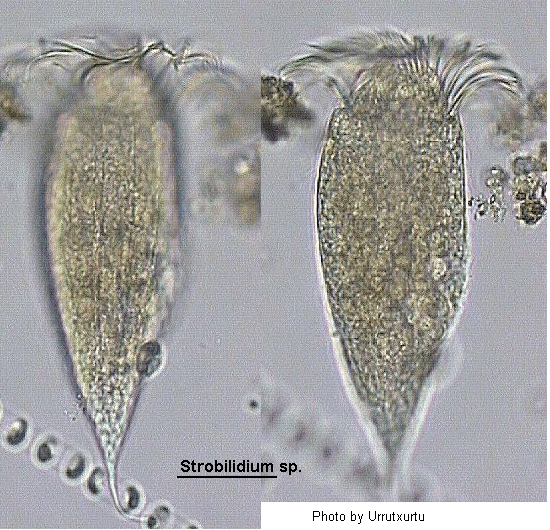
Class Polyhymenophora Jankowski, 1967: Subclass Spirotricha Butschli, 1889: Order Oligotrichida Butschli, 1887: Suborder Oligotrichina Butschli, 1887: Family Strobilidiidae Kahl in Doflein & Reichenow, 1929 (ref. ID; 4798)
Order Choreotrichida: Family Strobilidiidae (ref. ID; 7287, 7330) Synonym Stombilidium (ref. ID; 3389), Turbilina (ref. ID; 3389)
Strombilidium Schewiakoff, 1892 - Neave, 1940 (subsequent incorrect spelling) (ref. ID; 4356); Turbilina Enriques, 1908 (type species [by monotypy]: T. instabilis) (ref. ID; 4356) [ref. ID; 2014]
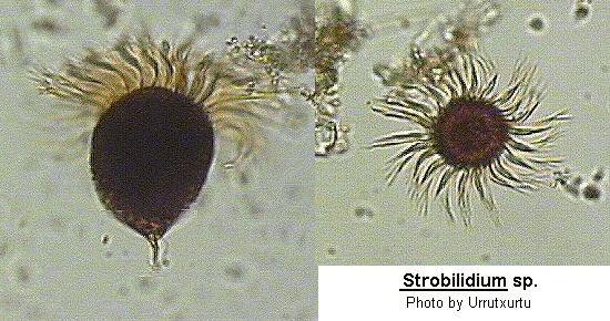
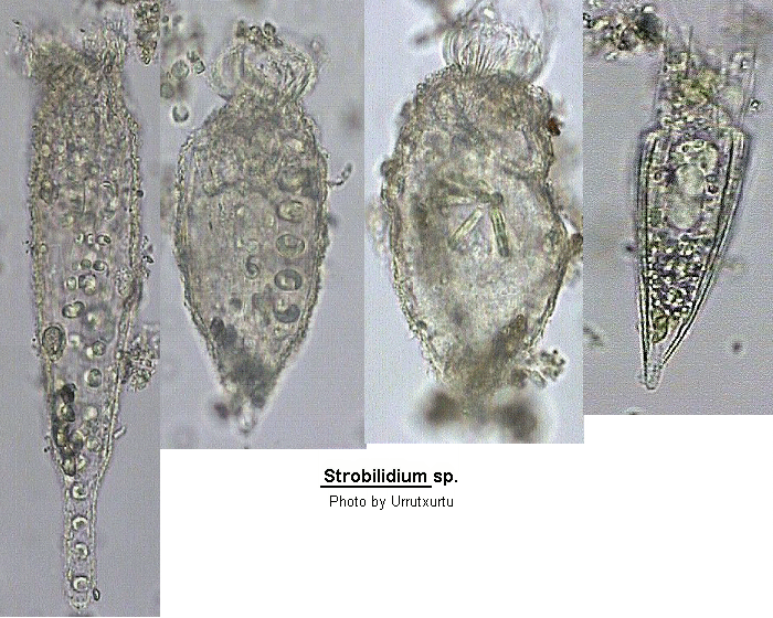
Spherical to elongate pyriform shape, swimming broad-end forward. The body is markedly twisted so that the somatic cilia when present spiral around the body. External ribs may be present in some species and these also spiral around the cell. When present, the somatic cilia are short, not very active and lie in 5 rows. In one species the terminal region of the cell is able to secrete a fragile mucilaginous thread by which if may attach itself to the substratum. The apical region of the cell carries a conspicuous AZM which is of the closed type. The original description did not include figures but a later paper contain figures and descriptions of 2 species.
Quote; Colin R. Curds, Michael A. Gates and David McL. Roberts "British and other freshwater ciliated protozoa Part II Ciliophora: Oligohymenophora and Polyhymenophora" Cambridge University Press, 1983 (ref. ID; 2014) [ref. ID; 3389]
Small to medium size species (12-170 um). Almost cylindrical in form; many species possess narrowed posterior region. Peristome is eccentrically placed in the apical area. There are somatic cirral rows in some species. Planktonic species in both marine and freshwater habitat. (ref. ID; 3389) [ref. ID; 4356]
Improved diagnosis; Strobilidiidae with several somatic kineties, some of which form a spiral at posterior end of cell. (ref. ID; 4356)
Remarks; Strobilidium is the type genus of the family Strobilidiidae, suborder Strobilidiina Jankowski, 1980, order Oligotrichida Butschli, 1887. Claparede & Lachmann (1859) used both spellings, Strombidion and Strombidium (in this order), when they established the genus. However, the spelling Strombidium succeeded. This can be justified with a very broad interpretation of Art. 24c and 32b of the International Code of Zoological Nomenclature (ICZM), saying that the correct spelling is that of the 1st reviser (ICZM, 1985), i.e. (Corliss 1961; Kahl 1932; Stein 1867). Thus, Strombidion should be considered a nomen oblitum and the correct spelling of the type species of Strobilidium is Strombidium caudatum. Lynn & Montagnes (1988) consider the arrangement of the somatic kineties to be an important generic criterion in strobilidiids. We agree and thus use the characteristic spiralling of the somatic kineties at the posterior a pole as genus character of Strobilidium. Strobilidiids lacking such a caudal spiral can be transferred to the ill-defined "desk-genus" Rimostrombidium Jankowski, 1978, for which the original definition (1978) is: "with ribbed cortex." Jankowski (1978) fixed as type Strobilidium velox Faure-Fremiet, 1924, which is rather similar to, e.g. Strobilidium lacustris Foissner et al., 1988. This kind of strobilidiid has slightly spiralling somatic kineties which do not extend to the posterior end of the cell, i.e. do not form a caudal spiral. Based on this improved definition, the following nominal species of Strobilidium are combined (in alphabetical order) with Rimostrombidium Jankowski: R. conicum (Kahl, 1932) n. comb.; R. epacrum (Lynn & Montagnes, 1988) n. comb.; R. humile (Penard, 1922) n. comb.; R. hyalinum (Mirabdullajev, 1985) n. comb.; R. kahli n. comb. (original nomenclatural act); R. lacustris (Foissner et al., 1988) n. comb.; R. lineolatum (Tucolesco, 1962) n. comb.; R. marinum (Faure-Fremiet, 1924) n. comb.; R. mirabile (Vuxanovici, 1962) n. comb.; R. multinucleatum (Lynn & Montagnes, 1988) n. comb.; R. polyhalinum (Tucolesco, 1962) n. comb.; R. saltans (Vuxanovici, 1962) n. comb.; R. sphaericum (Lynn & Montagnes, 1988) n. comb.; R. sulcatum (Tucolesco, 1962) n. comb. Several other poorly described species might also belong to Rimostrombidium (see Kahl 1932, Maeda 1986). (ref. ID; 4356)
Type species; Strombidium caudatum Fromental, 1876 (ref. ID; 4356)
- Strobilidium acuminatum (Faure-Fremiet, 1924) (ref. ID; 1621) or (Faure-Fremiet, 1924) Kahl, 1932 (ref. ID; 3389)
Syn; Strombidinopsis acuminatum Faure-Fremiet, 1924 (ref. ID; 3389) - Strobilidium adhaerens Schewiakoff, 1893 (ref. ID; 1621) or 1892 (ref. ID; 3389)
See; Strobilidium gyrans (ref. ID; 1621, 3389) - Strobilidium antarcticum (Busch) (ref. ID; 3544)
- Strobilidium armeniensis Zharikov, 1987
See; Rimostrombidium armeniensis (ref. ID; 4613) - Strobilidium caudatum (Fromental, 1874) Foissner, 1987 (ref. ID; 4730) or (Fromental, 1876) Foissner, 1987 (ref. ID; 4356, 4488, 4609)
Syn; Strobilidium adhaerens Schewiakoff, 1892 (ref. ID; 4356); Strobilidium caudatum (Fromental, 1874) Foissner, 1987 (ref. ID; 4356); nec; Strobilidium caudatum Kahl, 1932 (ref. ID; 4356); Strobilidium cometa (Muller, 1786) Dingfelder, 1962 (ref. ID; 4356); (?)Strobilidium gyrans Schewiakoff, 1893 - Deroux, 1974 (ref. ID; 4356); Strombidion caudatum Fromentel, 1874 (ref. ID; 4730) or 1876 (ref. ID; 4356, 4609); Strombidium claparedi Kent, 1881 (ref. ID; 4356, 4609); (?) Strombidium gyrans Stokes, 1887 (ref. ID; 4356, 4609, 4730); Strombidium gyrans Stokes var. transsylvanicum Lepsi, 1926 (ref. ID; 4356, 4609); Strombidium intermedium Maskell, 1887 (ref. ID; 4356, 4609); Strombidium velox Beardsley, 1902 (partim, Fig.5b) (ref. ID; 4356); Strombilidium gyrans Schewiakoff, 1893 - Fernandez-Leborans, 1983 (ref. ID; 4356); (?)Trichoda cometa Muller, 1773 (ref. ID; 4356); (?)Trichoda bomba Muller, 1773 (ref. ID; 4356); (?)Trichoda trochus Muller, 1786 (ref. ID; 4356); Turbilina instabilis Enriques, 1908 (ref. ID; 4356, 4609) - Strobilidium caudatum Kahl, 1932 (ref. ID; 3389) reported author and year? (ref. ID; 191, 1621, 1629)
- Strobilidium claparedei (Faure-Fremiet, 1924) nom. nov. (ref. ID; 3389)
Syn; Strombidinopsis claparedei Faure-Fremiet, 1924 (ref. ID; 3389) - Strobilidium conicum Kahl, 1932 (ref. ID; 3389, 3544, 4609) reported author and year? (ref. ID; 1621)
- Strobilidium diversum (Busch) (ref. ID; 3544)
- Strobilidium elegans Hada, 1970 (ref. ID; 3544 original paper)
See; Strombidium hadai (ref. ID; 3420) - Strobilidium elegans (Wulff, 1919) nom. nov. (ref. ID; 3389)
Syn; Lohmanniella elegans (Wulff, 1919) Kahl, 1932 (ref. ID; 3389); Sphaerotrichium elegans Wulff, 1919 (ref. ID; 3389) - Strobilidium elongatum (Leegaard) (ref. ID; 3544)
- Strobilidium epacrum (ref. ID; 1034, 1878)
- Strobilidium faurei Kahl, 1932
See; Parastrombidium faurei (ref. ID; 3389) - Strobilidium gyrans (Stokes, 1887) (ref. ID; 1219, 1335, 1621, 2245, 4671) reported year? (ref. ID; 1618, 3342, 3698) or (Stokes, 1887) Schewiakoff, 1892 (ref. ID; 3389) reported author and year? (ref. ID; 3954)
Syn; Strobilidium adhaerens Schewiakoff, 1893 (ref. ID; 1621) or 1892 (ref. ID; 3389); Strobilidium intermedium Maskell, 1888 (ref. ID; 1621); Strombidium caudatum Fromentel, 1876 (ref. ID; 1621); Strombidium claparedei Kent, 1882 (ref. ID; 1621, 3389); Strombidium gyrans Stokes, 1887 (ref. ID; 3389); Strombidium gyrans var. transsylvanicum Lepsi, 1926 (ref. ID; 3389); Strombidium intermedium Maskell, 1887 (ref. ID; 3389); Turbilina instabilis Enriques, 1908 (ref. ID; 3389) - Strobilidium hexakinetum (ref. ID; 7040)
- Strobilidium humile Penard, 1922 (ref. ID; 1621, 1629, 3389, 4609, 4613)
See; Rimostrombidium humile (ref. ID; 4613) - Strobilidium hyalinum Mirabdullaev, 1985 (ref. ID; 4613) reported author and year? (ref. ID; 4609)
See; Rimostrombidium hyalinum (ref. ID; 4613) - Strobilidium lacustris Foissner et al., 1988 (ref. ID; 1599 original paper)
See; Rimostrombidium lacustris (ref. ID; 4613) - Strobilidium lineolatum (Tucolesco, 1962) (ref. ID; 3389 Stribilidium misspelling?)
- Strobilidium marinum Faure-Fremiet, 1910 (ref. ID; 1621) or 1924 (ref. ID; 3388, 3389)
Syn; Strombidium marinum Faure-Fremiet, 1910 (ref. ID; 3389) - Strobilidium minimum (Gruber, 1884) (ref. ID; 1621) or (Gruber, 1884) Kahl, 1932 (ref. ID; 3389)
Syn; Arachnidium becheri Buddenbrock, 1920 (ref. ID; 3389); Strombidium minimum Gruber, 1884 (ref. ID; 3389) - Strobilidium mirabile Vuxanovici, 1962 (ref. ID; 3389)
- Strobilidium mucotectum (Busch, 1923) (ref. ID; 1621) or (Busch, 1924) Kahl, 1932 (ref. ID; 3389, 4917)
Syn; Strombidium mucotectum Busch, 1924 (ref. ID; 3389) - Strobilidium multinucleatum Lynn & Montagnes, 1988 (ref. ID; 7330)
- Strobilidium neptuni Montagnes & Taylor, 1994 (ref. ID; 7330 original paper)
- Strobilidium pelagicum Faure-Fremiet, 1924 (ref. ID; 1621, 3389)
Syn; Torquatella pelagica Faure-Fremiet, 1969 (ref. ID; 3389) - Strobilidium polyhalinum Tucolesco, 1962 (ref. ID; 3389)
- Strobilidium proboscidiferum (Milne, 1886) (ref. ID; 1621) or (Milne, 1886-7) Kahl, 1932 (ref. ID; 3389)
Syn; Strombidinopsis proboscidifer Milne, 1886-7 (ref. ID; 3389) - Strobilidium rapulum Yagiu, 1933
See; Strombidium rapulum (ref. ID; 3420) - Strobilidium saltans Vuxanovici, 1962 (ref. ID; 3389, 4609)
- Strobilidium sphaericum Lynn & Montagnes, 1988 (ref. ID; 7287, 7330)
- Strobilidium spiniferum (Leegaard, 1915) Kahl, 1932 (ref. ID; 3389)
Syn; Strombidium spiniferum Leegaard, 1915 (ref. ID; 3389) - Strobilidium spiralis (Leegaard, 1915) Lynn & Montagnes, 1988 (ref. ID; 7287, 7330) reported author and year? (ref. ID; 1878, 4371)
Syn; Lohmaniella spiralis (ref. ID; 1878) - Strobilidium striatum (Meunier, 1910) nom. nov. (ref. ID; 3389) or (Busch) (ref. ID; 3544)
Syn; Conocylis striata Meunier, 1910 (ref. ID; 3389); Strombidium striatum (Meunier, 1910) Kahl, 1932 (ref. ID; 3389) - Strobilidium sulcatum Claparede & Lachmann (ref. ID; 3544)
- Strobilidium sulcatum Tucolesco, 1962 (ref. ID; 3389)
- Strobilidium syowaensis Hada, 1970 (ref. ID; 3544 original paper)
See; Strombidium syowaensis (ref. ID; 3420) - Strobilidium tonsuratum (Meunier, 1910) nom. nov. (ref. ID; 3389)
Syn; Cephalotrichium tonsuratum Meunier, 1910 (ref. ID; 3389) - Strobilidium typicum Faure-Fremiet, 1924 (ref. ID; 1621) or (Lankester, 1874) Faure-Fremiet, 1924 (ref. ID; 3389)
Syn; Torquatella typica Lankester, 1874 (ref. ID; 3389) - Strobilidium undinum Martin & Montagnes, 1993 (ref. ID; 7287 original paper, 7330)
- Strobilidium velox Faure-Fremiet, 1924 (ref. ID; 1033, 1621, 3389, 4798)
See; Rimostrombidium velox (ref. ID; 4613) - Strobilidium veniliae Montagnes & Taylor, 1994 (ref. ID; 7330 original paper)
- Strombilidium viride Stein, 1867 (ref. ID; 4609)
Strobilidium acuminatum (Faure-Fremiet, 1924) (ref. ID; 1621) or (Faure-Fremiet, 1924) Kahl, 1932 (ref. ID; 3389)
Synonym
Strombidinopsis acuminatum Faure-Fremiet, 1924 (ref. ID; 3389)Descriptions
The body is slim, truncated anteriorly and pointed posteriorly. The numbers of adoral membranelles, 15-16, correspond to those of somatic cirral rows. Two oval macronuclei are present. Somewhat tiny substances accumulate at the posterior area. Swim very fast in a straight line. Frequently found in the pelagic region of the sea. (ref. ID; 3389)Measurements
Size, 110-115 um. (ref. ID; 3389)Strobilidium antarcticum (Busch) (ref. ID; 3544)
Descriptions
The ellipsoidal species with a rounded antapical end has a wide band composed of several short columns and slight oblique striae on the surface of the posterior half of the body. (ref. ID; 3544)Measurements
Length 60; breadth 40 um. (ref. ID; 3544)Strobilidium caudatum (Fromental, 1874) Foissner, 1987 (ref. ID; 4730) or (Fromental, 1876) Foissner, 1987 (ref. ID; 4356, 4488, 4609)
Synonym
Strobilidium adhaerens Schewiakoff, 1892 (ref. ID; 4356); Strobilidium caudatum (Fromental, 1874) Foissner, 1987 (ref. ID; 4356); nec; Strobilidium caudatum Kahl, 1932 (ref. ID; 4356); Strobilidium cometa (Muller, 1786) Dingfelder, 1962 (ref. ID; 4356); (?)Strobilidium gyrans Schewiakoff, 1893-Deroux, 1974 (ref. ID; 4356); Strombidion caudatum Fromentel, 1876 (ref. ID; 4356, 4609); Strombidium claparedi Kent, 1881 (ref. ID; 4356, 4609); (?)Strombidium gyrans Stokes, 1887 (ref. ID; 4356, 4609, 4730); Strombidium gyrans Stokes var. transsylvanicum Lepsi, 1926 (ref. ID; 4356, 4609); Strombidium intermedium Maskell, 1887 (ref. ID; 4356, 4609); Strombidium velox Beardsley, 1902 (partim, Fig.5b) (ref. ID; 4356); Strombilidium gyrans Schewiakoff, 1893 - Fernandez-Leborans, 1983 (ref. ID; 4356); (?)Trichoda cometa Muller, 1773 (ref. ID; 4356); (?)Trichoda bomba Muller, 1773 (ref. ID; 4356); (?)Trichoda trochus Muller, 1786 (ref. ID; 4356); Turbilina instabilis Enriques, 1908 (ref. ID; 4356, 4609) There is some confusion about the taxonomy of S. caudatum (Foissner, 1987), Dingfelder (1962) synonymized S. gyrans with Trichoda cometa. This is uncertain because shape (spherical) and movement (not fast) of T. cometa differ from S. caudatum. The movement and shape of T. trochus and S. caudatum are quite similar (O.F. Muller 1773, 1786). However, T. trochus and T. bomba are questionable synonyms for Halteria verrucosa (Fromental, 1876). These three poorly described ciliates are best considered nomina dubia. The outline of Strombidium velox is very similar to S. caudatum. In addition, S. velox forms a thread with which it fixes to the substrate and on which it gyrates (Beardsley 1902). However, Beardsley's Fig.5a (1902), which clearly shows a Strombidium, indicates that he mixed two differ species. Strobilidium caudatum Kahl, 1932 is certainly a different species. As a secondary homonym of S. caudatum (Fromental, 1876) Foissner, 1987, it has to be renamed; Strobilidium kahli nom. n. The other synonymy is according to (Kahl 1932; Maeda 1986). (ref. ID; 4356)Redescription
In vivo 50-65x40-50 um (average 56+/-6.7 x 42+/-5.0 um, n=8). Turbinate to inverted pyriform, slightly asymmetric, posterior end tapering and obliquely truncate, transverse section circular. Macronucleus horse shoe-shaped, close beneath zone of external adoral membranelles (external polykinetids), with opening in cytopharyngeal region. One spherical micronucleus adjacent to a macronucleus (rarely stained with protargol; position probably variable). Contractile vacuole at left side in posterior third of cell. Feeds on diatoms and flagellates. Movement very rapid, rotating about main body axis. When undisturbed for a longer while, cell secretes a thin mucous thread at caudal end ("scopula"), often very long (Grim & Halcrow 1979), and attaches to a substrate. Thread hardly recognizable unless small particles adhere to it. Frequently, cells swing on thread ("stalk") in a waving motion, still rotating. Disturbed individuals detach immediately and dart away. Somatic kineties restricted to posterior half of cell, composed of closely spaced cilia, very likely monokinetids. On ventral side usually two, rarely three, shortened somatic kineties which do not reach posterior pole; remaining kineties extend to caudal end where they form a distinct spiral. Cilia directed to left, 2-3 um long, shortened stubs in anterior portion of kineties often separated by small gap. Proximal third of cilia covered by cytoplasmic flap. External adoral membranelles on continuous cytoplasmic ridge; zone therefore appears as closed circle in top view, as stated previously (Corliss 1979; Deroux 1974; Faure-Fremiet 1970; Small & Lynn 1985). However, 4-6 increasingly elongated external membranelles and two very short internal adoral membranelles (internal polykinetids) extend into oral cavity, clearly showing that external membranelles form a spiral. In scanning electron micrographs, the external adoral membranelles are usually in furrows, which are, however, probably produced by a slight contraction of the oral ridge as we could not recognize them in living specimens. External membranelles composed of two long (ca. 10 um) and one shorter kinety (ca. 8 um), cilia about 25 um long and only proximally fused. Internal membranelles each consist of two rows of basal bodies. Anterior surface (peristomial bottom) slightly vaulted, with very short, funnel-shaped, acentric oral cavity defining ventral side and containing above mentioned elongated external and very small internal membranelles as well as single-rowed paroral membrane extending to center of anterior surface. A long bundle of pharyngeal fibers extends to posterior portion of cell. (ref. ID; 4356)Comments
The behavior of S. caudatum and the structure of the somatic kineties are well comparable with data by Grim & Halcrow (1979), i.e. cells attach with a mucous thread to the substrate, disturbed specimens leave their thread immediately, the somatic ciliature consists of short cilia which are proximally covered by a cytoplasm flap, and three somatic kineties form a distinct spiral at the posterior end of the cell. We did, however, not observe mucous material at the posterior pole. Strobilidum caudatum has been repeatedly investigated, but only some redescriptions are based on silver impregnation (Deroux 1974; Fernandez-Leborans 1983). However, the populations these authors studied differ from our isolate in some features, indicating that they might not be conspecific. The population of Deroux (1974) differs markedly from our S. caudatum by its occurrence (brackish water), by some morphologic characters (three of the five somatic kineties extend almost to the external adoral membranelles; adoral zone separated head-like from postoral portion), and by the site of the formation of the oral primordium (close beneath zone of external membranelles). Fernandez-Leborans' (1983) specimens differ by the larger size (in vivo 85-90 um) and by the higher number (29-30) and greater length (14 um) of the external membranelles; whether the four short, three-rowed "buccal membranelles" of this population are part of the elongated external membranelles or are internal membranelles cannot be seen from the figures provided. Further populations of S. caudatum should be studied to elucidate the identity of the isolates studied by Deroux (1974) and Fernandez-Leborans (1983). (ref. ID; 4356)Strobilidium caudatum Kahl, 1932 (ref. ID; 3389) reported author and year? (ref. ID; 191, 1621, 1629)
Descriptions
Shape is slender with a pointed posterior area. Kahl (1932) and Faure-Fremiet (1969) suggested this species resembles Strobilidium velox. Six ridges in the dorso-ventral area are spiralled. Found frequently in brackish water and occasionally in salt water near Kiel. (ref. ID; 3389)Measurements
Size, 50-70 um. (ref. ID; 3389)Strobilidium claparedei (Faure-Fremiet, 1924) nom. nov. (ref. ID; 3389)
Synonym
Strombidinopsis claparedei Faure-Fremiet, 1924 (ref. ID; 3389)Descriptions
The body is cylindrical or cylindro-conical. The anterior extremity is clearly truncated, the posterior end rounded. The peristomial area is confined at the apical zone where the circlet of adoral membranelles is present. The numbers of somatic cirral rows, which possess very short cirri, correspond to those of adoral membranelles. There are two macronuclei and one micronucleus. A contractile vacuole is situated near the buccal cavity. A freshwater species. (ref. ID; 3389)Comments
Claparede & Lachmann (1858, Page 372) described an animal, size 100 um in length, 30 um in width, which possessed an eccentrically placed buccal cavity and the spiralled apical membranelles. That species seems to be very similar to S. claparedei. (ref. ID; 3389)Measurements
Size, 70-80 um. (ref. ID; 3389)Strobilidium conicum Kahl, 1932 (ref. ID; 3389, 3544, 4609) reported author and year? (ref. ID; 1621)
Descriptions
The middle of the body is widened and the surface bears shallow furrows. The macronucleus is "horse-shoe" shaped. It rotates to advance, swings to and for and suddenly dashes forward over a long distance. Frequently found in brackish water. (ref. ID; 3389)The posterior part of the body is conical with distinct oblique striae. The macronucleus is oblong, and contractile vacuoles are usually visible. (ref. ID; 3544)
Measurements
Size, 25-40 um. (ref. ID; 3389)Length 30-55; breadth 22-32 um. (ref. ID; 3544)
Strobilidium diversum (Busch) (ref. ID; 3544)
Descriptions
The small form without striae is variable in form, and has a collaring band. (ref. ID; 3544)Measurements
Length 10-25; breadth 10-25 um. (ref. ID; 3544)Strobilidium elegans Hada, 1970 (ref. ID; 3544 original paper)
See
Strombidium hadai (ref. ID; 3420)Descriptions
The new form is small and slender, being more or less swollen at the middle and highly conical in the posterior half with a somewhat acute antapial end. The surface of the body is provided with a number (10-13) of longitudinal striae. The macronucleus is ovoidal near the center. (ref. ID; 3544)Comments
The new species is similar to S. elongatum (Leegaard) in general contour, but it is distinguishable from the latter in smaller size, slender form, and having a conical acute antapical end. (ref. ID; 3544)Measurements
Length 25-45; breadth 12-15 um. (ref. ID; 3544)Strobilidium elegans (Wulff, 1919) nom. nov. (ref. ID; 3389)
Synonym
Lohmanniella elegans (Wulff, 1919) Kahl, 1932 (ref. ID; 3389); Sphaerotrichium elegans Wulff, 1919 (ref. ID; 3389)Descriptions
The body is entirely globular. The circlet of frayed adoral membranelles is slightly spiraled and closed. The oval macronucleus contains many chromatin droplets. A marine species. (ref. ID; 3389)Comments
Wulff (1919) defined a new genus, Sphaerotichium, with this animal which as frayed apical membranelles. Kahl (1932) transferred it to the genus Lohmanniella. Reexamination of the original diagram and description suggest it should be included in the genus Strobilidium because of the arrangement of the apical membranelles. Strombidium sp. as described by Meunier (1910) is quite similar to this animal. (ref. ID; 3389)Measurements
Size, 17-30 um. (ref. ID; 3389)Strobilidium elongatum (Leegaard) (ref. ID; 3544)
Descriptions
The elongate form is fusiform with a slightly curved posterior part, and has a number of longitudinal striae on the surface. (ref. ID; 3544)Measurements
Length 60-150; breadth 27-77 um. (ref. ID; 3544)Strobilidium gyrans (Stokes, 1887) (ref. ID; 1219, 1335, 1621, 2245, 4671) reported year? (ref. ID; 1618, 3342, 3698) or (Stokes, 1887) Schewiakoff, 1892 (ref. ID; 3389) reported author and year? (ref. ID; 3954)
Synonym
Strobilidium adhaerens Schewiakoff, 1893 (ref. ID; 1621) or 1892 (ref. ID; 3389); Strobilidium intermedium Maskell, 1888 (ref. ID; 1621); Strombidium caudatum Fromentel, 1876 (ref. ID; 1621); Strombidium claparedei Kent, 1882 (ref. ID; 1621, 3389); Strombidium gyrans Stokes, 1887 (ref. ID; 3389); Strombidium gyrans var. transsylvanicum Lepsi, 1926 (ref. ID; 3389); Strombidium intermedium Maskell, 1887 (ref. ID; 3389); Turbilina instabilis Enriques, 1908 (ref. ID; 3389)Descriptions
Lateral border with rounded elevation near anterior end, posterior end truncate; in standing fresh water. (ref. ID; 1618)The body is pyriform, truncated anteriorly and the posterior area is narrowed. At the posterior extremity a mucus thread "scopula", which is used to attach to the substratum, is present. Deroux (1974) examined the detailed features of the arrangement of somatic cirral rows. Among the fine, oblique, somatic kineties the first three extend from near the adoral zone to the "scopula" in a spiralled row. The last two kineties are shorter than other three. The somatic cirri are not long. The "horse-shoe" shaped macronucleus is situated below the peristomial area with a micronucleus. A contractile vacuole is present posteriorly. When attached to the substratum by the "scopula" the animal has a waving motion, otherwise it swims very fast. Feeds on small green algae and diatoms. Widely spread and distributed in a shallow freshwater habitats. (ref. ID; 3389)
Comments
This animal was originally described as Strombidium gyrans by Stokes (1887). Schewiakoff (1892) constructed the new genus Strobilidium and described this species as Strobilidium adhaerens. Roux (1901) reviewed this animal and designated it Strobilidium gyrans Stokes using the diagram and description of Schewiakoff (1893). Penard (1922) named it Strobilidium gyrans (Stokes) Schewiakoff, 1893. (ref. ID; 3389)Measurements
40-70 um long. (ref. ID; 1618)54-47 um. (ref. ID; 3342)
Size, 60 um and 35-70 um according to Schewiakoff (1892) and Penard (1922). (ref. ID; 3389)
Strobilidium humile Penard, 1922 (ref. ID; 1621, 1629, 3389, 4609, 4613)
See
Rimostrombidium humile (ref. ID; 4613)Descriptions
The transparent body is cylindrical, the anterior area being widened. There is no protuberance in the front field. The "horse-shoe" shaped macronucleus is situated at the anterior end and a contractile vacuole is present at the posterior. It rotates slowly on one spot, moves convulsively back and forth over a short and dashes suddenly. Found among freshwater detritus. (ref. ID; 3389)Measurements
Size, 20-22 um. (ref. ID; 3389)Strobilidium lacustris Foissner et al., 1988 (ref. ID; 1599 original paper)
See
Rimostrombidium lacustris (ref. ID; 4613)Diagnosis
In vivo about 70-100x50-70 um (n=3); body cone-like, posterior region narrowed with a short spine-like projection; 9-10 equally long and equally spaced, right-spiraling somatic kineties; 31-32 adoral membranelles; single, horse-shoe-shaped macronucleus and single, lens-like micronucleus. (ref. ID; 1599)Descriptions
Body shape; rarely individuals observed with slightly turned-down collar. Transverse section round with small bulges where somatic kineties located. Caudal spine inconspicuous but present in all individuals. Pellicle thin, very fragile; most individuals burst at contact with cover glass. Macronucleus beneath the collar, filled with many small and large chromatin bodies (nucleoli?), with centers impregnating with protargol more weakly than peripheries; its "arms" membrane the cytopharynx. Micronucleus conspicuously large, situated in small indentation on dorsal side of macronucleus. Contractile vacuole at posterior end of body, just above caudal spine, with two inconspicuous canals. Cytoplasm colorless, but often appearing green from ingested algae. Somatic kineties running along small ribs, ending 1/5 body length from posterior pole, composed of short (ca. 2 um), immobile cilia, parallel to the body surface and close together, with fine projections which may contact pellicle. Adoral membranelles very conspicuous, encircle in an angle of about 45 degrees clockwise the frontal collar, which is more distinct in protargol-impregnated than in living cells. Pharynx located eccentrically, large, with long fibers reaching posterior region of cell. Each collar membranelle and first buccal membranelle composed of three long (ca. 17 um) rows kinetosomes bearing long (ca. 40 um) cilia apparently fused except for their distal ends; thus, distal parts of the membranelles appear frayed. Two shortened buccal membranelles, the innermost ca. 5 um long and composed of only two rows of kinetosomes. (ref. ID; 1599)Comments
Superficially, S. lacustris looks like S. caudatum (=S. gyrans). There are, however, distinct differences: S. lacustris never adheres to a substrate and never forms a stalk, which is so typical for S. caudatum. The somatic kineties are equally long in S. lacustris, whereas two of them are shortened in S. caudatum. As concerns the number of kineties and adoral membranelles, S. lacustris is very near to S. velox Faure-Fremiet, 1924. This species, however, measures only 30-70 um, has symbiotic green algae, and is pellicle is distinctly ribbed by the somatic kineties, which is never the case in S. lacustris. The pellicle of S. velox of Grim looks rather smooth, suggesting that it might be S. lacustris. The pattern of the adoral membranelles of S. lacustris fits exactly that described for S. velox, with the exception of the shortening of the buccal membranelles, which might have been overlooked. A paroral membrane, which is present in other strobilids, has not been observed in S. lacustris; it may not have impregnated or may be inconspicuous. (ref. ID; 1599)Type locality
Epilimnion of Lake Svinsjoen near Oslo, Norway. (ref. ID; 1599)Strobilidium lineolatum (Tucolesco, 1962) (ref. ID; 3389 Stribilidium misspelling?)
Descriptions
The transparent body is spherical and furnished with 10-12 striations in the somatic area. Between the striation the pellicle is slightly protuberant. Found in underground water in a cave. (ref. ID; 3389)Measurements
Size, 15 um. (ref. ID; 3389)Strobilidium marinum Faure-Fremiet, 1910 (ref. ID; 1621) or 1924 (ref. ID; 3388, 3389)
Synonym
Strombidium marinum Faure-Fremiet, 1910 (ref. ID; 3389)Descriptions
The thin apical collar is distinct erectile. Below the apical collar there is a deep constriction. The posterior extremity is sharply pointed. Thirty to thirty two apical membranelles are present. Paroral membrane is present. Somatic cirral rows are dispersed and not distinct. The macronucleus is spherical with a micronucleus. Movement is very fast following a corkscrew path. A marine pelagic species. (ref. ID; 3389)Measurements
Size, 100 um. (ref. ID; 3389)Strobilidium minimum (Gruber, 1884) (ref. ID; 1621) or (Gruber, 1884) Kahl, 1932 (ref. ID; 3389)
Synonym
Arachnidium becheri Buddenbrock, 1920 (ref. ID; 3389); Strombidium minimum Gruber, 1884 (ref. ID; 3389)Descriptions
The body shape is variable, usually oval, rounded posteriorly. The anterior extremity is triangularly truncated and adoral membranelles are frayed according to Kahl (1932). Small particles, such as sand grains, often adhere to the gelatinous materials which covers almost all the body. Two macronuclei are present, although Kahl (1932) once observed the animal with only one macronucleus. The animal shows a bouncingly and/or rotatively movement with occasional pause on the detritus. A marine species, especially common in eutrophic area. (ref. ID; 3389)Measurements
Size, 30 um and 30-60 um according to Gruber (1884) and Kahl (1932), respectively. (ref. ID; 3389)Strobilidium mirabile Vuxanovici, 1962 (ref. ID; 3389)
Descriptions
The body shape is like Strobilidium humile, but this animal is 5-6 times bigger. Apical membranelles are 40 um in length. The macronucleus is ellipsoidal. A contractile vacuole is present laterally. The animal moves quickly in a straight line followed by rapid rotation and a short pause. Found in freshwater in Bucharest. (ref. ID; 3389)Measurements
Size, 105 um. (ref. ID; 3389)Strobilidium mucotectum (Busch, 1923) (ref. ID; 1621) or (Busch, 1924) Kahl, 1932 (ref. ID; 3389, 4917)
Synonym
Strombidium mucotectum Busch, 1924 (ref. ID; 3389)Descriptions
The body is cylindrical. The posterior area is three sided and shortly pointed. A pellicle is present under a layer of gelatinous material, 3.5 um in thickness which covers the body. Many small nuclei and large oil droplets are present. A marine species found in Java. (ref. ID; 3389)Measurements
Size, 135 um. (ref. ID; 3389)Strobilidium neptuni Montagnes & Taylor, 1994 (ref. ID; 7330 original paper)
Diagnosis
Cell shape, subspherical, although, in fixed samples, flattened of one side due to the characteristic structure of kinety 2. Cell 40 um (range, 33-57) long and 45 um (38-55) wide. External polykinetidal zone (EPZ) composed of 38 (36-40) external polykinetids (EPk). Cilia of the EPk are short near the cell centre and increase in length with distance from the cell centre (minimum length 3-5 um, maximum length ~30 um). Additionally, every second cilium is ~5 um shorter than those adjacent to it. Internal polykinetidal zone (IPZ) composed of 13 (9-20) internal polykinetids (IPk). IPk and EPk usually contriguous but those further from the cytostome may be continuous. The IPZ sites above an acentrically placed deprresion that leads to the cytostome. Cytopharyngeal fibers present. Paroral kinety begins parallel to the last IPk (near the cytostome) and lies internal to the EPZ. Cilia of the paroral kinety are ~10-15 um long and lie on the oral surface inside the region defined by the EPZ. Five somatic kineties (K) present, whose cilia (2-3 um long) are directed to the right (when viewed aborally). The somatic kineties are partially covered by a cytoplasmic flap. K1 is slightly and dextrally spiralled (viewed aborally) and extends from the posterior of the cell to just below the EPZ. K2 describes an arc that encloses a space between K1 and K2 by extending from the posterior of the cell to a position half way along K1; at this point K2 reflects away from K1 and produces a small (2-4 um) "crook". K3 and K4 are simple; they originate perpendicular to and near the aboral third of K2 and extend anteriorly to just below the EPZ. K5 originates one-third of the cell length from the aboral end and runs anteriorly, paralleling K1. One C-shaped macronucleus, positioned anteriorly around the oral cavity and below the EPZ, has its opening near the cytostome. The micronuclei (not always visible) are indented into the macronucleus 180 degrees around from the cytostome. Cell surface often covered by 2-4 um rods (possibly extrusomes), which can strain darkly with protargol. (ref. ID; 7330)Discussion of related species
The family Strobilidiidae sensu stricto exhibits the following characters: somatic kineties with short cilia overlain by a cytoplasmic flap and a closed circle of external polykinetids; the genus Strobilidium possesses these some characters (Lynn and Montagnes 1988). Following these criteria Lynn and Montagnes (1988) made Lohmanniella spiralis Leegaard, 1915 (a commonly reffered to marine ciliate) a junior synonym for Strobilidium spiralis. [The ciliate described above (Strobilidium spiralis) exhibits kineties whose cilia are directed to the right and covered by a cytoplasmic flap. They are therefore, similar in general structure to those of S. gyrans (see Deroux, 1974) and other Strobilidium species described above. We consider this a diagnostic character for the genus Strobilidium]. More recently, Petz and Foissner (1992) have placed a number of species of Strobilidium in the genus Rimostrombidium Jankowski, 1978: [Lynn and Montagnes consider the arrangement of the somatic kineties to be an important generic criterion in strobilidiids. We agree and thus use the characteristic spiralling of the somatic kineties at the posterior pole (of S. gyrans) as a genus characters of Strobilidium. Strobilidiids lacking such a caudal spiral can be transferred to the ill defined "desk-genus" Rimostrombidium Jankowski, 1978...] In Lynn and Montagnes (1988) and in Montagnes and Lynn (1991), differences in kinetal structure (e.g. cytoplasm flap and mono- vs. dikinetids) are used to diagnose genera within the family Strobilidiidae. They then use the arrangement (e.g. length and position) of the somatic kineties to make more subtle specific distinctions within the genera. The unstated logic of this is that kinetal structure is likely conservative, while kinetal arrangement, although distinctive, is more likely to show convergence. Thus, the suggestion of Petz and Foissner (1992), to transfer many species of Strobilidium to the "desk-genus" Rimostrombidium, based on a distinction of kinetal arrangement, is in conflict with the criteria proposed by Lynn and Montagnes (1988) and is based on potentially unsound characters. Furthermore, resurrection of the "desk-genus" Rimostrombidium for this group creates unnecessary etymological confusion, as it implies phylogenetic affinity to the genus Strombidium, which is distinctly different from any of the strobilidiids (Montagnes and Lynn 1991; Petz and Foissner 1992; Small and Lynn 1985). We therefore have maintained the genus Strobilidium for this and other identifications, following Lynn and Montagnes (1988). Strain IA is identical to Strobilidium spiralis (Leegaard, 1915) Lynn and Montagnes, 1988 except for two features: 1) S. spiralis has only one micronucleus indented into the macronucleus (180 degrees around from the cytostome), while strain IA has two, and 2) K5 on S. spiralis originates anteriorly, extends posteriorly, and then curves anteriorly around to describe a partial circle; but K5 on strain IA originates one-third of the cell length from the aboral end and runs anteriorly, parallelling K1. These two differences provide distinctions between the two species. (ref. ID; 7330)Remarks
The swimming behaviour of this ciliate is typical of many strobilidiids; it rotates in one position in the water, with its anterior end facing outward. This swimming pattern is punctuated by "jumps" of two to five cells lengths. Such jumps are stimulated by agitation of the culture and by the presence of other moving objects (e.g. a pipette tip) but may also be stimulated by changes in food concentrations (unpubl. data). (ref. ID; 7330)Etymology
The secific epithet, neptuni, stems from Neptune, the Latin name for the god of the sea: "the Governour of the Sea, the Father of the Rivers and the Fountains, and the Son of Saturn by Ops." (ref. ID; 7330)Time and locality of isolation
Early March, Indian Arm, British Columbia, Canada (122 degrees 53'W, 49 degrees 21'N), at a depth of 2 m, temperature of 8 degrees C, and salinity of 16 0/00. (ref. ID; 7330)Deposition of type material
A slide, USNM #47748, of protargol-stained cells representing an hapantotype from strain IA resides in the Ciliate Type Specimen Collection, US Natural History Museum, Smithsonian Institution, Washigton, D.C. (ref. ID; 7330)Strobilidium pelagicum Faure-Fremiet, 1924 (ref. ID; 1621, 3389)
Synonym
Torquatella pelagica Faure-Fremiet, 1969 (ref. ID; 3389)Descriptions
The body is roundly widened at the anterior area and proportionally narrowed to the posterior area. The striped somatic region possesses no ciliature. The pellicle is very soft. It has fibrous endoplasm containing large oil droplets. Two oval macronuclei are present. It feeds on Peridinium. A marine species found in pelagic coastal water. (ref. ID; 3389)Measurements
Size, 135-170 um and 220 um during cell division. (ref. ID; 3389)Strobilidium polyhalinum Tucolesco, 1962 (ref. ID; 3389)
Descriptions
The body shape is an inverted cone and it is dark brown or brownish orange in colour. The adoral membranelles are confined to the anterior zone and form a circle. Quite frequently found in Lake Tekirghiol where the concentration of sulphate is high. (ref. ID; 3389)Measurements
Size, 12-15 um. (ref. ID; 3389)Strobilidium proboscidiferum (Milne, 1886) (ref. ID; 1621) or (Milne, 1886-7) Kahl, 1932 (ref. ID; 3389)
Synonym
Strombidinopsis proboscidifer Milne, 1886-7 (ref. ID; 3389)Descriptions
The body has an elastic pellicle and is globular in shape, truncated anteriorly. Inside the circlet of apical membranelles, there is a membrane-like collar. An elastic and retractile protuberance, which is located inside this collar, can be stretched higher than the collar or drawn back entirely below the collar. In the somatic area, deep longitudinal grooves and short cirri are present. The body is usually full of large refractive particles. The macronucleus is elongate and placed transversely below the apical protuberance. A contractile vacuole is present in the posterior region. A marine species. (ref. ID; 3389)Measurements
Size, 36 um. (ref. ID; 3389)Strobilidium saltans Vuxanovici, 1962 (ref. ID; 3389, 4609)
Descriptions
The body, shaped like an inverted cone, is truncated anteriorly. The somatic area is striated with 14-16 stripes. The macronucleus is a "horse-shoe" shape. The animal moves very quickly with short breaks. Found in lake water in Bucharest. (ref. ID; 3389)Measurements
Size, 28 um. (ref. ID; 3389)Strobilidium spiniferum (Leegaard, 1915) Kahl, 1932 (ref. ID; 3389)
Synonym
Strombidium spiniferum Leegaard, 1915 (ref. ID; 3389)Descriptions
Described and named using a fixed specimen. The smooth body is widened in the middle and pointed posteriorly. The nucleus is spherical. Found in the North Sea. (ref. ID; 3389)Measurements
Size, around 70 um. (ref. ID; 3389)Strobilidium striatum (Meunier, 1910) nom. nov. (ref. ID; 3389) or (Busch) (ref. ID; 3544)
Synonym
Conocylis striata Meunier, 1910 (ref. ID; 3389); Strombidium striatum (Meunier, 1910) Kahl, 1932 (ref. ID; 3389)Descriptions
The body is elongated and turbinatus form. Somatic area displays distinct stripes with broad furrows of variable shape and number. The macronucleus is ovoid. (ref. ID; 3389)The body is fusiform and usually twice as long as broad. Several striae are on the surface of the posterior conical part of the body. (ref. ID; 3544)
Comments
Busch's (1930) Strombidium striatum may be a different species from this animal. (ref. ID; 3389)Measurements
Size, 70-90 um according to Kahl (1932). (ref. ID; 3389)Length 25-50; breadth 18-27 um. (ref. ID; 3544)
Strobilidium sulcatum Claparede & Lachmann (ref. ID; 3544)
Descriptions
The marked characteristics of the species common in fresh water, is having a wide band composed of short striae in the posterior half of the body. (ref. ID; 3544)Measurements
Length 35-37; breadth 28-30 um. (ref. ID; 3544)Strobilidium sulcatum Tucolesco, 1962 (ref. ID; 3389)
Descriptions
The body is dorso-ventrally flattened and the posterior area is acuminated. There are deep furrows in the somatic area. Movement is rapid. A marine species. (ref. ID; 3389)Measurements
Size, 25 um. (ref. ID; 3389)Strobilidium syowaensis Hada, 1970 (ref. ID; 3544 original paper)
See
Strombidium syowaensis (ref. ID; 3420)Descriptions
The body is large and ovoidal, and consists of a widely conical anterior crown with well-developed membranelles, a median band figured with inverted triangles on the surface, and a roundly conical posterior region containing numerous granules. A contractile vacuole usually appears. (ref. ID; 3544)Comments
The new species is different from the known forms of Strobilidium in having the median characteristic band. (ref. ID; 3544)Measurements
Length 82-160; breadth 67-90 um. (ref. ID; 3544)Strobilidium tonsuratum (Meunier, 1910) nom. nov. (ref. ID; 3389)
Synonym
Cephalotrichium tonsuratum Meunier, 1910 (ref. ID; 3389)Descriptions
Ovoid in shape, posterior area narrowed. Membranelles of the adoral zone are "lamella-like". There is no ciliation in the somatic area. The pellicle is thin but the sarcoplasm is thick and only slightly transparent. The macronucleus is large and elongated, containing dense granules. Described from fixed specimens collected from seawater. (ref. ID; 3389)Comments
Meunier (1910) showed two kinds of diagram, one showing the opened circlet of apical membranelles and the other closed. Kahl (1932) and Faure-Fremiet (1969) designated this animal in the Families Strobilidiidae and Halteriidae, respectively. In this work it had been tentatively kept in the genus Strobilidium because of the absence of somatic ciliature. (ref. ID; 3389)Measurements
Size, 100 um. (ref. ID; 3389)Strobilidium typicum Faure-Fremiet, 1924 (ref. ID; 1621) or (Lankester, 1874) Faure-Fremiet, 1924 (ref. ID; 3389)
Synonym
Torquatella typica Lankester, 1874 (ref. ID; 3389)Descriptions
The body is widely round posteriorly and anterior area is elongate. The membranelles of the adoral zone, which surround the depressed peristomial area, beat powerfully and there is a short paroral membrane in the buccal cavity. A fine pellicle covers the whole body, where somatic ciliature is absent. Two ovoid macronuclei with two associated micronuclei are present in the central area and a contractile vacuole is located at posteriorly. Lankester (1874) found this animal in a saprobic habitat, but Faure-Fremiet (1924) isolated it in the pelagic sea. (ref. ID; 3389)Measurements
Size, 80 um. (ref. ID; 3389)Strobilidium undinum Martin & Montagnes, 1993 (ref. ID; 7287 original paper, 7330)
Diagnosis
The following diagnosis is based on observations of over 30 protargol-stained specimens from Indian Arm, B.C. Five (4-6) internal and 23 (21-24) external polykinetids. Six ciliated somatic kineties, of 15-20 monokinetids, radiating from near posterior pole. One C-shaped macronucleus positioned anteriorly around oral cavity and below external polykinetid zone. (ref. ID; 7287)Descriptions
Cell subspherical, 21 um (16-29) long and 17 um (15-23) wide. Internal polykinetid zone lies in an acentric pit that leads to cytostome. Internal polykinetids and external polykinetids usually continuous although furthest 2-3 from cytostome may be continuous. Cilia of somatic kineties directed to right when viewed aborally. (ref. ID; 7287)Generic position and comparison to related species
Strobilidium undinum that continuous kineties with cilia directed to the right, suggesting they are partially covered by a cytoplasmic flap. However, a flap was not observed as this structure can be difficult to discern by protargol staining. Still, we believe a flap was present, placing it in the genus Strobilidium. The U-shaped macronucleus and cell shape of Strobilidium undinum are similar to Strobilidium sphaericum Lynn and Montagnes, 1988, and S. spiralis (Leegaard, 1915) Lynn and Montagnes, 1988. However, differences exist in somatic ciliature, oral membranelles and cell size: S. sphaericum has more somatic kineties than S. undinum (10 vs. 6) and is longer (46 um vs. 21 um). Strobilidium undinum has a similar number of external polykinetids as S. sphaericum (26 vs. 23); however, those of the latter spiral into a central depression that leads to the cytostome. In addition, S. sphaericum lacks an internal polykinetid zone; S. spiralis possesses only five somatic kineties, which do not radiate in the manner described for S. undinum; and S. spiralis is longer than S. undinum (36 um vs. 21 um), and contains more membranelles within the external and internal polykinetid zones (26 externals and 13 internals vs. 23 externals and 5 internals, respectively). (ref. ID; 7287)Etymology
The specific eipthet, undinum, stems from the Latin root undin, meaning spirit of the waves. (ref. ID; 7287)Type locality
Shallow marine waters (top 5 m; salinity of 22-25 0/00) of Indian Arm, B.C., Canada (latitude 49 degrees 22'N, longitude 122 degrees 55'W), taken February 1990. (ref. ID; 7287)Deposition of type material
Holotype and paratype specimens of Strobilidium undinum have been marked on a slide of protargol-impregnated cells (from this field study) and has been deposited in the Ciliate Type Specimen Slide Collection, USNM #43112 and #43136, respectively, U.S. Natural History Museum, Smithsonian Institution, Washigton, DC, USA. (ref. ID; 7287)Strobilidium velox Faure-Fremiet, 1924 (ref. ID; 1033, 1621, 3389, 4798)
See
Rimostrombidium velox (ref. ID; 4613)Descriptions
[ref. ID; 1033]This isolates is ca. 69 um long and has 11 body kineties which are ca. 60% of the body length. The posterior end is distinctly pointed. The somatic kineties contain very short cilia that are very close together and lie partially beneath a thin fold of plasma membrane and cytoplasm. In a recent taxonomic guide (Maeda 1986) this species was described as not having somatic kineties. They are present in my strain; however, the cilia often are not visible, even in protargol preparations. Oral ciliature includes a peristomial AZM (not in the buccal cavity) of ca. 26 membranelles (polykinetids) and a single paroral membrane. Two of the membranelles and the paroral membrane continue into the buccal cavity. The outer limiting membrane appears to be poorly fixed. This membrane (of the body and cilia) has never been seen as a trilaminar structure. This is probably due to poor fixation. An inner membrane, alveoli, and epiplasm also have not been seen. (ref. ID; 1033)