Urocentrum
Urocentrum Nitzsch, 1827 (ref. ID; 2014)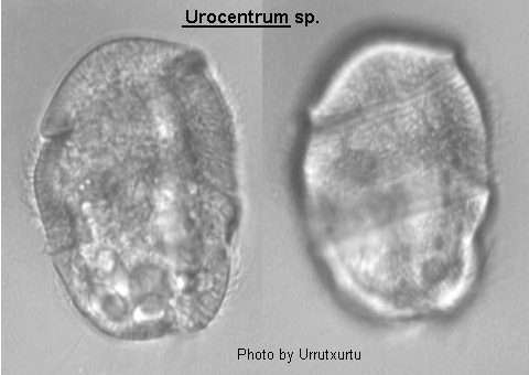
Medium-sized (80-120 um long) barrel-shaped ciliate with broadly rounded ends and slightly waisted equatorially, narrowing slightly posteriorly. Oral aperture equatorial, located at anterior end of a longitudinal groove situated in the posterior body half, oval in outline with its axis lying obliquely to that of the major body axis. There is undulating membrane on the right edge of the aperture and 3 membranelles within the cavity. The posterior oral groove is ciliated and towards the posteriorly. Somatic ciliation is restricted to 3 girdles, (1) around the equator consisting of short dense cilia; (2) immediately anterior to this a much wider girdle of longer less densely packed cilia which may or may not reach the body apex and (3) a girdle lying in the posterior body half and consisting of long less-dense cilia which never cover the terminal pole. Contractile vacuole posterior, fed by 4 long serving canals. Macronucleus horseshoe-shaped lying in a transverse plane in posterior body half surrounding micronucleus. Single species genus.
Quote; Colin R. Curds, Michael A. Gates and David McL. Roberts "British and other freshwater ciliated protozoa Part II Ciliophora: Oligohymenophora and Polyhymenophora" Cambridge University Press, 1983 (ref. ID; 2014) [ref. ID; 4716]
The genus Urocentrum has been considered till now as the single member of the Family Urocentridae included in the Suborder Peniculina (Corliss 1979; Levine et al. 1980). Levine et al. (1980) point out the suitability of promoting this suborder to the systematic category of Order on the basis of the conclusions given by the French authors (Puytorac and Grain 1976; Puytorac et al. 1974). Lynn (1979) reaches the same conclusion using the numerical phenetic analysis of 57 cortical characters. Recently Puytorac et al. (1984) reattempted to classify the phylum Ciliophora applying the phenetic analysis of 122 different features and to mention the Order Peniculida although they exclude the genus Urocentrum from it and transferred it to the Order Hymenostomatida as a member of the new Suborder Turaniellina. According to this paper the Order Hymenostomatida includes three Suborders, Tetrahymenina, Ophryoglenina and Turaniellina with two Families: Turaniellidae and Urocentridae. The data used by these French authors to classify the genus Urocentrum have been furnished with the papers of Roque (1961, 1973) and Didier (1971). We have already expressed our reasons for disagreement on a number of points with the conclusions reached by these authors, specially in those features related to stomatogenesis. Despite of the similarity of the characteristics of the somatic cortex in Turaniella and in Urocentrum and their approximation to those in tetrahymenines rather than those in peniculines, stomatogenesis is very different in both species. Turaniella presents a parakinetal stomatogenesis as in tetrahymenines and the oral structures of the opisthe derive from one or more somatic kineties of the parental cell. On the contrary, Urocentrum exhibits a buccokinetal type of stomatogenesis pattern, typical of peniculines. Therefore we conclude that the position of U. turbo in the new Suborder Turaniellina (Order Hymenostomatida) should be reconsidered, while the genus Urocentrum should remain in the Order Peniculida. (ref. ID; 4716)
- Urocentrum turbo (O.F. Muller) (ref. ID; 1618, 3342, 4716), (O.F. Muller, 1786) Kahl, 1931 (ref. ID; 1219, 1622, 1629, 2245) or (O.F. Muller, 1786) Nitzsch, 1827 (ref. ID; 4611) reported author and year? (ref. ID; 191, 3292)
Syn; Calceolus cypripedium Diesing, 1866 (ref. ID; 1622); Cercaria turbo O.F. Muller, 1786 (ref. ID; 1622, 4611); Peridinopsis cyripedium J. Clark, 1866 (ref. ID; 1622); Urocentrum trichocystus Smith, 1897 (ref. ID; 1622)
Urocentrum turbo (O.F. Muller) (ref. ID; 1618, 3342, 4716), (O.F. Muller, 1786) Kahl, 1931 (ref. ID; 1219, 1622, 1629, 2245) or (O.F. Muller, 1786) Nitzsch, 1827 (ref. ID; 4611) reported author and year? (ref. ID; 191, 3292)
Synonym
Calceolus cypripedium Diesing, 1866 (ref. ID; 1622); Cercaria turbo O.F. Muller, 1786 (ref. ID; 1622, 4611); Peridinopsis cyripedium J. Clark, 1866 (ref. ID; 1622); Urocentrum trichocystus Smith, 1897 (ref. ID; 1622)Descriptions
Body cylindrical and ventrally slightly flattened; constricted at the middle; 2 broad girdles of cilia and 1 eccentric posterior tuft; a zone of short cilia in the constricted area; buccal cavity subequatorial with 1 membrane and 2 short undulating membranelles; in the postoral area, a longitudinal zone with small cilia; macronucleus horseshoe-shaped and posteriorly located; a single micronucleus; contractile vacuole terminal, and with 8 collecting canals; large number of trichocysts all over the body. (ref. ID; 1219) [ref. ID; 4716]Urocentrum turbo is cylindrically shaped with middle third of the body slightly pinched in. The nuclear apparatus is situated in the posterior third of the cell and it consists of a horseshoe-shaped macronucleus with rounded and more voluminous extremes and a spherical micronucleus located next to the narrowest point of the macronucleus. (ref. ID; 4716)
- Stages 1. Rearrangement of the kinetosomes of the oral field into three segments, each with two rows of kinetosomes, following a gradient from the zone closest to the paroral and from the anterior part to the posterior one of the buccal cavity. These three segments represent the anterior part of each of the three peniculi primordia.
- Stage 2. The paroral kinety doubles and the oral field breaks into two part as a result of the rearragement of its kinetosomes: the anterior part, designated right area of the oral field (RFA) which has fewer kinetosomes, is close to the anterior exremes of the three peniculi primordia and the second on reffered to as the left area of the oral field (LFA), presents a more posterior position in the oral cavity.
- Stage 3. The number of rows of the penicular primordia increases so that each of them contains 3 rows of kinetosomes. At the same time, these primordia grow longer due to the addition of groups of three kinetosomes which come from the proliferation of the kinetosomes in the LFA.
- Stage 4. The kinetosomes of the RFA proliferate and then a double row of kinetosomes appears in the anterior part of this area, and forms the paroral kinety of the opisthe. Simultaneously to the above the primordia of the peniculi go on increasing in length by proliferation. These peniculi are the result of the configuration of kinetosomes in groups of three which then migrated from the LFA. The paroral kinety of the proter, which was previously doubled, increases its number of kinetosomes to produce the oral field of the proter.
- Stage 5. The growth in length of the three peniculi stops and the LFA disappears. That is, the oral infraciliature of the opisthe is completely differentiated.
- Stage 6. The P1 and P2 of the proter appear one after the other. At the same time, the oral structures of the proter and those of the opisthe progressively move away. The posterior parts of the oral structures of both the proter and opisthe then move downwards, to enter the body on the right side and the oral cavities of both cells are formed in this way. These cavity are antero-posteriorly oblique to the longitudinal axis of the body. (ref. ID; 4716)
Measurements
Length 50-80 um. (ref. ID; 1219, 1618)82 um. (ref. ID; 3342)