Stylonychia
Stylonychia Ehrenberg, 1830 (ref. ID; 2014)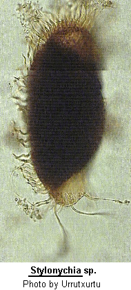

Family Oxytrichidae Ehrenberg, 1838: Subfamily Stylonychinae Berger & Foissner, 1997 (ref. ID; 4894 subfamily original paper) Synonym Kerona O.F. Muller, 1786 (ref. ID; 2014); Prosopsenus Andre, 1916 (ref. ID; 2014); Stylonichia (ref. ID; 2014) [ref. ID; 593]
Of all Stylonychia species, which have been described only 3 species have a length of more than 150 um. Stylonychia mytilus, Stylonychia grandis, Stylonychia kahli. (ref. ID; 593) [ref. ID; 2014]
Inflexible, elongate, oval, dorso-ventrally flattened body with a large and powerful AZM supported anteriorly by a collar. 2 rows of marginal cirri not continuous posteriorly, but 3 long, strong and prominent caudal cirri present. The genus has the typical fronto-ventral and transverse arrangement of cirri with 3 strong anterior frontal cirri. The dorsal surface has several longitudinal rows of short cilia. Macronucleus in two parts each with an adjacent micronucleus. Several species have been recorded.
Quote; Colin R. Curds, Michael A. Gates and David McL. Roberts "British and other freshwater ciliated protozoa Part II Ciliophora: Oligohymenophora and Polyhymenophora" Cambridge University Press, 1983 (ref. ID; 2014) [ref. ID; 4894]
Improved characterization; Undulating membranes in Stylonychia pattern. One right and one left row of marginal cirri, distinctly separate posteriorly. Six dorsal kineties. Caudal cirri present and often distinctly elongated. Primordium II of the proter originates from oral primordium. Primordium V and VI of the proter originate from frontoventral cirrus IV/3 or from postoral ventral cirrus V/4. Primordium IV of the opisthe originates from postoral ventral cirrus V/4. Dorsal morphogenesis in Oxytricha pattern. (ref. ID; 4894)
Remarks; Morphogenetic and molecular biological data (Schlegel & Steinbruck 1986) suggest that Stylonychia is not monophyletic. However, molecular biological data on the closely related genera Steinia and Coniculostomum should be awaited before splitting. In vivo, small species, for instance the very common S. pustulata, are easily confused with Oxytricha spp., which have, however, a more slender and flexible body and intersecting undulating membranes. (ref. ID; 4894)
Type species (subsequent designation by Fromentel 1875); Trichoda mytilus Muller, 1773 (ref. ID; 4894)
- Stylonychia appendiculata Ehrenberg, 1838
See; Diophrys appendiculata (ref. ID; 4408) - Sytlonychia bifaria (ref. ID; 2857)
- Stylonychia curvata (ref. ID; 1621)
- Stylonychia fissiseta Claparede & Lachmann, 1858 (ref. ID; 1621, 4150)
- Stylonychia grandis (ref. ID; 191, 593)
- Stylonychia grandis Hemberger, 1982 (ref. ID; 4719)
- Stylonychia grandis Maupas (ref. ID; 1621)
- Stylonychia histrio (ref. ID; 191)
- Stylonychia kahli (ref. ID; 593)
- Stylonychia lanceolata Ehrenberg, 1838
See; Pleurotricha lanceolata (ref. ID; 4121) - Stylonychia lemnae Ammermann & Schlegel, 1983 (ref. ID; 7423)
- Stylonychia lemnae Steinbruck & Schlegel, 1983 (ref. ID; 593, 4078) reported author and year? (ref. ID; 191, 3649, 4471, 4719)
See; Stylonychia mytilus Ehrenberg (ref. ID; 4078) - Stylonychia macrostyla Sterki, 1878 (ref. ID; 1621)
- Stylonychia muscorum Kahl, 1932 (ref. ID; 4609) reported author and year? (ref. ID; 1621)
See; Stylonychia stylomuscrom (ref. ID; 4609) - Stylonychia mytilus-complex (ref. ID; 4609)
Syn; Trichoda mytilus O.F. Muller, 1773 (ref. ID; 4609) - Stylonychia mytilus (ref. ID; 191, 475, 3627, 3628, 3751, 3936, 4379, 4380, 4381, 4471, 4719, 7592)
- Stylonychia mytilus Ehrenberg, 1838 (ref. ID; 593, 662, 1621, 1629, 1896, 2245, 3116, 4078) or 1938 (ref. ID; 3979) reported year? (ref. ID; 646, 1219, 3342, 3698)
- Stylonychia mytilus Foissner et al., 1991 (ref. ID; 4488)
- Stylonychia mytilus (O.F. Muller) (ref. ID; 1618), (Muller, 1773) Ehrenberg, 1830 (ref. ID; 7423)
- Stylonychia notophora Stokes, 1885 (ref. ID; 1621, 4150) reported year? (ref. ID; 1618, 3342) reported author and year? (ref. ID; 191, 4471)
- Stylonychia ovalis Dragesco, 1966 (ref. ID; 4150)
See; Stylonychia vorax (ref. ID; 4609) - Stylonychia pusilla (ref. ID; 1621, 4471)
Syn; Stylonychia mytilus var. pusilla Sterki, 1878 (ref. ID; 1621) - Stylonychia pustulata Ehrenberg (ref. ID; 1308, 1618, 1621, 1629, 2245), (O.F. Muller, 1786) Ehrenberg, 1835 (ref. ID; 4488, 4609, 7423), (O.F. Muller, 1786) Ehrenberg, 1838 (ref. ID; 4150) reported author and year? (ref. ID; 191, 2857, 4375, 4471)
See; Clara pustulata (ref. D; 7423)
Syn; Kerona pustulata O.F. Muller, 1786 (ref. ID; 4609) - Stylonychia pustulata Wirnsberger et al., 1985 (ref. ID; 4719)
- Stylonychia putrina Stokes, 1885 (ref. ID; 1621, 1629, 2245, 4609) reported year? (ref. ID; 1219, 1618, 5462) reported author and year? (ref. ID; 191)
- Stylonychia stylomuscorum Foissner, Blatterer, Berger & Kohmann, 1991 (ref. ID; 4609) reported author and year? (ref. ID; 1629)
Syn; Stylonychia muscorum Kahl, 1932 (ref. ID; 4609) - Stylonychia vorax Stokes, 1885 (ref. ID; 4150, 4488, 4609, 7423) reported author and year? (ref. ID; 191, 1629, 2857, 4471)
See; Clara vorax (ref. D; 7423)
Syn; Stylonychia ovalis Dragesco, 1966 (ref. ID; 4609) - Stylonychia vorax Wirnsberger et al., 1985 (ref. ID; 4719)
Stylonychia grandis Maupas (ref. ID; 1621)
Descriptions
This species is the same size as S. mytilus but has a macronucleus consisting of four parts. Borror (1972) does not list this species as a Stylonychia. (ref. ID; 593)Stylonychia kahli (ref. ID; 593)
Descriptions
This species has a length of 200 um. The rows of right and left marginal cirri are, however, confluent posteriorly, and therefore we believe that this species does not belong to the genus Stylonychia. (ref. ID; 593)Stylonychia lemnae Steinbruck & Schlegel, 1983 (ref. ID; 593, 4078) reported author and year? (ref. ID; 191, 3649, 4471, 4719)
See
Stylonychia mytilus Ehrenberg (ref. ID; 4078)Descriptions
(Formerly called Stylonychia mytilus, Variety I, Ammermann). The shape of the cells distinguishes this species from Stylonychia mytilus. The right and left margins of the cells are nearly parallel. The ciliature of the cell does not differ from the typical pattern. The macronucleus consists of two parts, which are connected by a fine process. Usually 2-3 micronuclei are also present. (ref. ID; 593)Comments
See Stylonychia mytilus. (ref. ID; 4719)Etymology
S. lemnae (like S. mytilus) is often found in ponds which are covered with Lemna minor (Arales, Lemnaceae). Terra typica: Pond IV of the "Spitzberg forest" (48 degrees 31'N, 9 degrees 02'E) near Tuebingen. This pond lies 436 m above sea level. Some slides of S. lemnae from this pond will be sent to the Smithsonian Institute, Washington, D.C., U.S.A. (ref. ID; 593)Measurements
230+/-12 um after starvation for one day, approximately 270 um before division, approximately 120 um minimal length (after 3 days of starvation). (ref. ID; 593)Stylonychia mytilus (ref. ID; 191, 475, 3627, 3628, 3751, 3936, 4379, 4380, 4381, 4471, 4719, 7592)
Descriptions
The body measures 250-300 um in length and is dorsoventrally flattened, with a rounded anterior end and a slightly pointed posterior end. S. mytilus has two macronuclei with a size of 41x16 um on average and two spherical micronuclei. The somatic ciliature consists of 8 frontal cirri, 5 ventral cirri, 5 transverse cirri, 3 caudal cirri, 20-25 right marginal cirri and 15 left marginal cirri (Tuffrau 1965). The AZM has 65-80 membranelles and there are two "undulating membrane" (paroral and endoral). (ref. ID; 475)Conjugation. (ref. ID; 7592)
Comments
[ref. ID; 4719]- Stomatogenesis. Small groups of basal bodies evolve very close to the 1st (and sometimes the 2nd too) uppermost transverse cirrus, which remains unchanged. The number of basal bodies increases and forms a longish anarchic field. Only the 1st third of the parental undulating membranes is reorganized, because no further dispersal of basal occurs. The primordium for the undulating membranes of the opisthe evolves from the oral anarchich field. At this time also the first new adoral membranelles arrange themselves.(ref. ID; 4719)
- Development of the cirral primordia. In the opisthe the frontal-ventral-transverse (FVT) anlagen 1, 2, and 3 originate from the oral primordium. The anlagen 4, 5, and 6 are produced by the right postoral ventral cirrus. In the proter anlage 1 evolves from the parental undulating membranes; anlage 2 from the oral primordium of the opisthe; anlage 3 from the left posterior frontal cirrus; anlage 4 from the right posterior frontal cirrus; and the anlagen 5 and 6 originate from the right postoral ventral cirrus. Uniformly 6 FVT-anlagen are produced in each filial product. The development of the marginal primordia always starts within the right row and proceeds more rapidly there than in the left row. Later the events in the marginal rows appear to be correlated. Five frontal cirri, the buccal cirrus, the left postoral ventral cirrus, the posterior ventral and transverse cirri, and several marginal cirri do no participate in the formation of primordia. The left posterior frontal cirrus and the right postoral ventral cirrus proliferate basal bodies and are partly dissolved. The rest is resorbed in the last stages of the division, together with the other cirri, which are not involved in building the anlagen. (ref. ID; 4719)
- Further events. The nuclear apparatus divides in the same manner as described in Stylonychia mytilus by Wallengren (1900) and Frick (1967, 1968). The differentiation of new cirri -the FVT-anlagen 1-6 always generate 1, 3, 3, 3, 4, and 4 cirri-, their migration, and the development of the dorsal primordia are indistinguishable from those of other members of the genus Stylonychia (Hemberger 1982; Wirnsberger et al. 1985). (ref. ID; 4719)
Stylonychia mytilus Ehrenberg, 1838 (ref. ID; 593, 662, 1621, 1629, 1896, 2245, 3116, 4078) or 1938 (ref. ID; 3979) reported year? (ref. ID; 646, 1219, 3342, 3698)
Sibling species
Formerly named S. mytilus Variety II; Ammermann, 1965. (ref. ID; 593)Originally, Stylonychia mytilus was defined as a single species; however, a more detailed genetic and morphological analysis has shown that this "species" consists of two sibling species: S. mytilus and S. lemnae (formerly variety I and II) (Ammermann et al., 1983). (ref. ID; 4078)
Descriptions
The shape is typical, more pronounced in starved than in well fed animals; while the right and left margins of the cells are nearly parallel in S. lemnae, in S. mytilus the left margin (from dorsal) has a bulge in the region of the peristome. This typical cell shape is pictured in Ehrenberg's original description. (ref. ID; 593)The dorsal side bears only small cilia (sensory bristles); the ventral side is furnished with strong cilia or cirri arranged in characteristic rows and groups; rows of cirri near both right and left margins, interrupted posteriorly; 8 strong frontal cirri, 5 ventral ones behind the buccal area, 5 transverse cirri, and 3 caudal ones; the caudal cirri show fringes ate the end. Buccal area furnished with conspicuous adoral membranelles, and 1 long undulating membrane at the right side, the cytostome is located at the posterior end of the buccal area; 2 macronuclei and 2 micronuclei; 1 contractile vacuole near the posterior end of the buccal area. The size of Stylonychia may vary considerable; furthermore, it should be stressed that the family Oxytrichidae as well as the genus Stylonychia itself includes a great number of species, some of which may rather closely resemble the one described here. All identification features should thus be checked carefully. (ref. ID; 1219)
Measurements
Length 100-300 um. (ref. ID; 1219)200 um. (ref. ID; 3342)
Stylonychia mytilus (O.F. Muller) (ref. ID; 1618), (Muller, 1773) Ehrenberg, 1830 (ref. ID; 7423)
Descriptions
Fresh, brackish and salt water. (ref. ID; 1618)Measurements
100-300 um long. (ref. ID; 1618)Stylonychia notophora Stokes, 1885 (ref. ID; 1621, 4150) reported year? (ref. ID; 1618, 3342) reported author and year? (ref. ID; 191, 4471)
Descriptions
Standing water. (ref. ID; 1618)Measurements
About 125 um long. (ref. ID; 1618)100-115x34-50 um. (ref. ID; 3342)
Stylonychia pustulata Ehrenberg (ref. ID; 1308, 1618, 1621, 1629, 2245), (O.F. Muller, 1786) Ehrenberg, 1835 (ref. ID; 4488, 4609, 7423) or (O.F. Muller, 1786) Ehrenberg, 1838 (ref. ID; 4150) reported author and year? (ref. ID; 191, 2857, 4375, 4471)
See
Clara pustulata (ref. D; 7423)Synonym
Kerona pustulata O.F. Muller, 1786 (ref. ID; 4609)Descriptions
Fresh water. (ref. ID; 1618)Body inflexible, elliptical, in vivo 48-124x26-83 um. Both ends rounded, margins more or less parallel. About 2:1 flattened dorso-ventrally, central part often very bulged. Macronuclear segments two, ovoid, in vivo 16x7 um, lying left of the median, filled with numerous little masses (average 0.8 um in diameter, n=12). Micronuclei two, in vivo 4x3 um, each in close contact with one of the macronuclear segments. Contractile vacuole, in vivo 10 um, on the left-hand border above the middle of the body. Endoplasm full of bright yellow, refringent inclusions, 2-3 um, of various forms. Food vacuoles, in vivo up to 32 um, include diatoms sometimes reaching one-third of the body length. Even at low magnification the specimens, except for the peristome field and the area around the transverse cirri, show a dark color. Found in company with algae, Stylonychia mytilus, Paramecium bursaria, and Coleps. Movement moderately slow, gliding, and often standing still for sometimes, rotation, around the longer axis on disturbance. Conjugation was observed very infrequently. Mixed clones in most cases encysted, but never showed pairing. Cysts have a toothed surface and a fine, bright endoplasm. Marginal cirri in vivo about 15 um long, all bases are nearly of the same size, 1.5x0.7 um, and equally spaced within the posteriorly clear-cut rows. Right marginal row begins at the level of the first adoral membranelles and is composed of many more cirri than the left one. Three anterior frontal cirri enlarged, in vivo 19-23 um long, bases about 2.6x2.6 um. Four posterior frontal cirri, arranged in an oblique hook-shaped row, and one enlarged buccal cirrus. Adoral zone of membranelles often longer than half of the body length. After protargol staining the bases of the longest membranelles measure 22 um. Undulating membranes straight and never overcrossing. Pharyngeal fibers distinguishable in vivo. Two postoral and three ventral cirri, from which the posteriormost one is adjacent to the right transverse cirrus. Transverse cirri five, in vivo 20-24 um long, characteristically arranged in a group of four cirri in an oblique row adjoined by the fifth transverse cirrus and the equal-sized ventral cirrus. All transverse cirri project beyond the posterior border. Six dorsal kineties. Four long rows composed of 19-29 pairs of basal bodies (average 26, n=5). Note that kinety 4 extends to the anterior end of the body, contrary to S. vorax. Two very short rows in the frontal area. Caudal cirri three, in vivo 16-25 um long, unfringed, very stiff, projecting laterally. The distance between the two right caudal cirri is always smaller than that between the second and third ones. (ref. ID; 4150)
Comments
Stylonychia pustulata was identified by the shape of the body, the arrangement and the number of cortical elements, the mode of moving, the dorsal-bristle complex, the toothed cysts, and the typical inclusions. We agree with Borror (1983) that S. fissiseta Claparede & Lachmann, 1858, which Hemberger (1981) transferred to S. mytilus, is a synonym of S. pustulata. Both consider S. notophora Stokes, 1885 to be indistinguishable from S. pustulata. We agree because of its morphological similarities and the de novo stomatogenesis, described in Sapra et al, 1970; however, some authors reported four micronuclei in this species. (ref. ID; 4150)Measurements
About 150 um long. (ref. ID; 1618)Stylonychia putrina Stokes, 1885 (ref. ID; 1621, 1629, 2245, 4609) reported year? (ref. ID; 1219, 1618, 5462) reported author and year? (ref. ID; 191)
Descriptions
Similar to S. mytilus but smaller, and right and left sides are more or less parallel; caudal cirri unfringed, 2-4 transverse cirri protruding slightly beyond the posterior end. (ref. ID; 1219)Measurements
Length 125-145 um. (ref. ID; 1219)125-150 um long. (ref. ID; 1618)