Opercularia
Opercularia Goldfuss, 1820 (ref. ID; 2014) or Stein, 1854 (ref. ID; 1555) reported year? (ref. ID; 1248, 1618)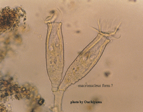
Family Epistylidae Kahl, 1935 (ref. ID; 1248) [ref. ID; 2014]
Colonial. Inverted bell-shaped zooids mounted upon branced non-contractile stalk. The peristome without a lip and its rim is often scalloped and rarely smooth. The macronucleus usually elongate, never rounded. But curved or C-shaped and held in a horizontal position. Often epizooic. The genus is easily mistaken for Orbopercularia which differs only by having a rounded macronucleus. It could also be confused with Epistlyis which has a definite peristomial lip.
Quote; Colin R. Curds, Michael A. Gates and David McL. Roberts "British and other freshwater ciliated protozoa Part II Ciliophora: Oligohymenophora and Polyhymenophora" Cambridge University Press, 1983 (ref. ID; 2014) [ref. ID; 1555]
Type species; Opercularia articulata (Ehrenberg) Stein, 1854 (ref. ID; 1555)
- Opercularia allensi Stokes, 1887 (ref. ID; 1557, 1620, 2245, 4610), allensii Stokes (ref. ID; 2861)
See; Opercularia nutans (ref. ID; 4610) - Opercularia arboricolum (Biegel, 1954) (ref. ID; 922) reported author and year? (ref. ID; 7614)
- Opercularia archiorbopercularia Foissner (ref. ID; 3698) reported author and year? (ref. ID; 1114)
- Opercularia articulata Ehrenberg, 1838 (ref. ID; 1114, 1620, 1629, 2573) reported year? (ref. ID; 3698) or (Ehrenberg) Stein, 1854 (ref. ID; 1555) reported author and year? (ref. ID; 3292)
- Operucularia articulate Goldfuss, 1820 (ref. ID; 4610)
Syn; Opercularia confusa Stiller, 1940 (ref. ID; 4610); Opercularia phryganeae Kahl, 1935 (ref. ID; 4610) - Opercularia asellicola Kahl, 1935 (ref. ID; 1248)
- Opercularia assellicola (ref. ID; 1620)
- Opercularia aymmetrica (ref. ID; 4610)
- Opercularia berberina Linnaus (ref. ID; 1620)
- Opercularia clepsinis Popow, 1904 (ref. ID; 1620)
- Opercularia coarctata (Claparede & Lachmann, 1858) (ref. ID; 1219, 1620, 1629, 2245, 3664, 7614) reported year? (ref. ID; 2861, 4735, 4759) or (Claparede & Lachmann, 1858) Roux, 1901 (ref. ID; 4610) reported author and year? (ref. ID; 191, 1555, 3689), cf. coarctata (ref. ID; 1557)
Syn; Epistylis coarctata Claparede & Lachmann, 1858 (ref. ID; 1113, 4610); Epistylis cylindrica Kusters, 1974 (ref. ID; 1113); Epistylis opercularia Stiller, 1942 (ref. ID; 1113); Opercularia arenicola Greeff, 1873 (ref. ID; 1113); Opercularia coarctata Roux, 1901 (ref. ID; 1113); Opercularia curvicaule Curds, 1964 (ref. ID; 1113); Opercularia fabrei Faure-Fremiet, 1905 (ref. ID; 1113); Opercularia frondicola Precht, 1936 (ref. ID; 1113); Opercularia gracilis Faure-Fremiet, 1904 (ref. ID; 1113, 1620); Opercularia halophila Kusters, 1974 (ref. ID; 1113); Opercularia microdiscum Faure-Fremiet, 1904 (ref. ID; 1113, 1620); Opercularia minima Kahl, 1935 (ref. ID; 1113, 1620); Opercularia reifi Matthes, 1950, 1951 (ref. ID; 1113); Opercularia sinuans Stiller, 1942 (ref. ID; 1113); Opercularia stammeri Matthes, 1950 (ref. ID; 1113); Rhabdostyla muscorum Kahl, 1935 (ref. ID; 1113) - Opercularia confusa Stiller, 1940 (ref. ID; 4610) reported year? (ref. ID; 2861)
See; Opercularia articulata Goldfuss, 1820 (ref. ID; 4610) - Opercularia collegata (ref. ID; 1555, 3689)
- Opercularia curvicaula (Penard) Curds (ref. ID; 2861)
Syn; Pyxidium curvicaula Penard (ref. ID; 2861) - Opercularia cylindrata Wrzesniowski, 1870 (ref. ID; 1620, 4488)
- Opercularia dimorpha (Dogiel & Furssenko, 1920) (ref. ID; 1555)
- Opercularia elongata Kellicott, 1884 (ref. ID; 1620)
- Opercularia epistyliformis (ref. ID; 1555, 3689)
- Opercularia eumelita (ref. ID; 3689)
- Opercularia faurei Collin, 1909 (ref. ID; 1620) reported author and year? (ref. ID; 3689)
- Opercularia glomerata Roux, 1901 (ref. ID; 1620)
- Opercularia halophila Kusters, 1774 (ref. ID; 4654 original paper)
- Opercularia henneguyi (Faure-Frem., 1905) (ref. ID; 1248)
Syn; Pyridium henneguyi (ref. ID; 1248) - Opercularia hustedti Sommer, 1951 (ref. ID; 1248 original paper)
- Opercularia irritabilis Hempel (ref. ID; 1620)
- Opercularia jumpi Guhl, 1972 (ref. ID; 7635 original paper)
- Opercularia laccobii Matthes & Guhl (ref. ID; 7619)
- Opercularia lichtensteini Stein, 1868 (ref. ID; 1248, 1620)
See; Orbopercularia sp. - Opercularia marceli (Remy, 1928) (ref. ID; 1555)
- Opercularia medians Collin, 1909 (ref. ID; 1620)
- Opercularia microdiscum Faure-Fremiet (ref. ID; 2861)
- Opercularia microstoma Stein, 1868 (ref. ID; 1620)
- Opercularia minima Kahl, 1935 (ref. ID; 1248) reported year? (ref. ID; 2861)
- Opercularia moldavica Sramek-Husek, 1948
See; Epistylis chrysemydis (ref. ID; 4610) - Opercularia nutans (Ehrenberg, 1838) (ref. ID; 1219, 1248, 1620, 1629, 2245) reported year? (ref. ID; 2861, 3343) or (Ehrenberg, 1831) Stein, 1854 (ref. ID; 4610)
Syn; Epistylis nutans Ehrenberg, 1831 (ref. ID; 4610); Opercularia allensi Stokes, 1887 (ref. ID; 4610) - Opercularia parallela Kirk, 1885 (ref. ID; 1620)
- Opercularia penardi Kahl, 1935 (ref. ID; 1248) reported author and year? (ref. ID; 1620)
- Opercularia phryganeae Kahl, 1935 (ref. ID; 4610) reported year? (ref. ID; 2861) reported author and year? (ref. ID; 1620)
See; Opercularia articulate Goldfuss, 1820 (ref. ID; 4610) - Opercularia plicatilis Stokes, 1884 (ref. ID; 1620) reported year? (ref. ID; 1618)
- Opercularia pseudoberberina (ref. ID; 4610)
- Opercularia ramosa Stokes (ref. ID; 1219, 1335)
- Opercularia rugosa Kellicott, 1884 (ref. ID; 1620)
- Opercularia stenostoma Stein-D'Udekem (ref. ID; 1618, 1620)
- Opercularia subcorethrae Sommer, 1951 (ref. ID; 1248 original paper)
- Opercularia venusta Foissner (ref. ID; 3698) reported author and year? (ref. ID; 1114)
Opercularia allensi Stokes, 1887 (ref. ID; 1557, 1620, 2245, 4610), allensii Stokes (ref. ID; 2861)
See
Opercularia nutans (ref. ID; 4610)Descriptions
O. allensii has zooids 60-140 um (in the average 100 um) and colonies up to 0.5 mm in diameter. In contracted state the individuals are hanging like at the precedent species. The stalks are longitudinally striated and irregularly segmented. The distal branches are relatively short. Catching bacteria, small algae and diatoms of the genus Navicula this species is common in spring and autumn in running and standing waters. It attaches to aquatic vegetation and easily also to the glass slides. (ref. ID; 2861)Opercularia coarctata (Claparede & Lachmann, 1858) (ref. ID; 1219, 1620, 1629, 2245, 3664, 7614) reported year? (ref. ID; 2861, 4735, 4759), (Claparede & Lachmann, 1858) Roux, 1901 (ref. ID; 4610) reported author and year? (ref. ID; 191, 1555, 3689), cf. coarctata (ref. ID; 1557)
Synonym
Epistylis coarctata Claparede & Lachmann, 1858 (ref. ID; 1113, 4610); Epistylis cylindrica Kusters, 1974 (ref. ID; 1113); Epistylis opercularia Stiller, 1942 (ref. ID; 1113); Opercularia arenicola Greeff, 1873 (ref. ID; 1113); Opercularia coarctata Roux, 1901 (ref. ID; 1113); Opercularia curvicaule Curds, 1964 (ref. ID; 1113); Opercularia fabrei Faure-Fremiet, 1905 (ref. ID; 1113); Opercularia frondicola Precht, 1936 (ref. ID; 1113); Opercularia gracilis Faure-Fremiet, 1904 (ref. ID; 1113, 1620); Opercularia halophila Kusters, 1974 (ref. ID; 1113); Opercularia microdiscum Faure-Fremiet, 1904 (ref. ID; 1113, 1620); Opercularia minima Kahl, 1935 (ref. ID; 1113, 1620); Opercularia reifi Matthes, 1950, 1951 (ref. ID; 1113); Opercularia sinuans Stiller, 1942 (ref. ID; 1113); Opercularia stammeri Matthes, 1950 (ref. ID; 1113); Rhabdostyla muscorum Kahl, 1935 (ref. ID; 1113); Telotrochidium johanninae Faure-Fremiet, 1950 (ref. ID; 1113)Descriptions
Colonies small, consisting of 3-6 individuals; body elongate; peristomal area small and not constricted form the body; buccal area with distinct oblique disk which is set off from the border by a deep incision; obvious undulating membrane. Macronucleus is horseshoe-like, its axis transverse to the longitudinal. (ref. ID; 1219)O. coarctata has zooids 45-65 um long and stalks reaching 50-100 um. The colonies consist of only 3-6 individuals. The stalk is unstriated, slender, sessile on the flocks of activated sludge or among other slime organisms. Body is elongate, peristomial area small and not constricted from the body. There is a deep incision between the oblique disc and the peristomial border. The undulating membrane can be easily seen. Macronucleus is of the horseshoe-type, its axis is transverse to the longitudinal axis of the zooid. The pellicle is generally smooth and may be striated close to the stalk. The species is common in alpha-mesosaprobical environment, often together with Vorticella putrina and V. convallaria: oxidation ponds, biofilters, activated sludge, where it may reach an abundancy of more than 100,000 individuals per 1 ml. Nusch (1970) observed in experiments with sewage and brewery wastes a little different variety with 4-10 zooids in a colony. (ref. ID; 2861) [ref. ID; 7614]
Oral structures:
Remarks
The oral infraciliature of Opercularia coarctata is very like to that of Opercularia arboricolum (Foissner, 1981). Guhl (1979) has considered these species as probable synonims whereas Foissner (1981) thinks that are separate species. (ref. ID; 7614)Examined material
O. coarctata was isolated from a pond water sample in Asturias (Spain). (ref. ID; 7614)Measurements
Individuals 45-65 um, stalk about 50-100 um. (ref. ID; 1219)Opercularia confusa Stiller, 1940 (ref. ID; 4610) reported year? (ref. ID; 2861)
See
Opercularia articulata Goldfuss, 1820 (ref. ID; 4610)Descriptions
O. confusa has zooids 85-90 um long and colonies consisting of 5-6 individuals. This species is living in the littoral zone of lakes, ponds and small water bodies on living as well as dead aquatic plants. The telotrochs leave the stalks without forming the aboral ring of cilia. The species can be classified as an indicator of natural decomposition processes common in all types of surface waters. (ref. ID; 2861)Opercularia curvicaula (Penard) Curds (ref. ID; 2861)
Synonym
Pyxidium curvicaula Penard (ref. ID; 2861)Descriptions
O. curvicaula resembles the species O. coarctata, but is smaller and slender. Zooids 40 up to 80 um, the stalk is inapparent, resembles a mycoidal hypha and supports a colony of up to 17 individuals. There are 3 whorls to the peristomial disc. The short macronucleus winds around the cytopharynx. The base of the body is finely striated but the rest of the pellicle is smooth. The food vacuoles are lemon-shaped. The species was originally described as Pyxidium curvicaule by Penard but redescribed by Curds (1964). In Great Britain Curds & Cockburn (1970) found it infrequently in biofilters and activated sludge. (ref. ID; 2861)Opercularia microdiscum Faure-Fremiet (ref. ID; 2861)
Descriptions
Zooids 70-90 um, abundant colonies up to 0.5 mm. The stalk is long, longitudinally striated, but not segments. The body is cylinder-like with a wide opening. The peristomial disc is borne upon a short process and is about one-fifth of the body width. The pellicle is striated and the macronucleus winds around the cytopharynx in the anterior region of the body. The single contractile vacuole opens into the wide pharynx. The number of food vacuoles and reserve granules is small. It was described as an epibiont on the larvae of Eristalis tenax living in highly polluted waters. Curds & Cockburn (1970) found it in biofilters, often is large numbers. (ref. ID; 2861)Opercularia minima Kahl, 1935 (ref. ID; 1248) reported year? (ref. ID; 2861)
Descriptions
O. minima has individuals 35-40 um on stalks which are much shorter than the zooids. Colonies consist of 2 or 4 individuals. The peristome disc is borne on a long process and the mouth region is quite wide. The pellicle is striated. There is one contractile vacuole. The macronucleus is situated in the upper half of the body and winds around the cytopharynx parallel to the peristome. The short stalk is longitudinally striated and segmented. The species was described as an ectocommensal on the legs of freshwater mites, but occurs also in moderate or small numbers in biofilters and activated sludge. (ref. ID; 2861)Opercularia nutans (Ehrenberg, 1838) (ref. ID; 1219, 1248, 1620, 1629, 2245) reported year? (ref. ID; 2861, 3343) or (Ehrenberg, 1831) Stein, 1854 (ref. ID; 4610)
Synonym
Epistylis nutans Ehrenberg, 1831 (ref. ID; 4610); Opercularia allensi Stokes, 1887 (ref. ID; 4610)Descriptions
Disk comparatively high and stalk more or less annulated (at least in the anterior region). (ref. ID; 1219)O. nutans has zooids 60-140 um which are erect when whirling bur declined (hanging) when contracted. Abundant colonies reach up to 3 mm in size. The stalk is long, more or less annulated, mainly in the anterior region. The disc is comparative high. This species is attached to freshwater algae (Cladophora, Enteromorpha). To stones, insect larvae and easily attaches to submerged glass slides and can be classified as beta-mesosaprobic with inclination to alpha-mesosaprobity. It is one of the best and easily recognizable indicators of pollution of rivers and artificial reservoirs. (ref. ID; 2861)
Measurements
Individuals 60-141 um, colonies up to 3 mm. (ref. ID; 1219)Length of cell 60 um. (ref. ID; 3343)