The World of Protozoa, Rotifera, Nematoda and Oligochaeta
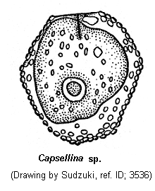
Capsellina
Capsellina Penard, 1909 (ref. ID; 7582)
Quote from ref. ID; 3536
Class Flosea (Leidy, 1879): Order Gromiida (Claparede & Lachmann, 1859): Family Gromiidae (ref. ID; 7582)
[ref. ID; 1618]
Test hyaline, ovoid, membranous; with or without a second outer covering; aperture long slit; a single nucleus; one or more contractile vacuoles; filose pseudopodia; fresh water. (ref. ID; 1618)
[ref. ID; 1923]
Shell ovoid, membranous, hyaline or brown. Aperture linear. (ref. ID; 1923)
[ref. ID; 7582]
The shell of testate amoebae, dorso-ventrally squeezed, is most often elliptic through the major longitudinal axis and for some rhizopods only, nearly circular. The posterior pole is regularly rounded, while for the opposit one, two convex half circles end tangentially to the median plane, on the level of the pseudostome. The shell wall is thin, flexible, transparent, withocet any ornamentation and completely filled with the protoplasm. Through the aperture, of which the true shape is estimated in light microscopy with difficulty, protrudes a very narrow pseudopodial trunk which opens in a typical filosa-like pseudopodial apparatus. Completely hyaline, this apparatus shows:
- slime-ended pseudopodia which break like a bayonnet, while striking,
- hyaline and utmost thin lamina, never thick-set, nor rounded as the lobosa type.
These two filopodial features can be isolated or associated: In this case, the hyalin lamina unites the slim-ended pseudopodia as a webbed-foot. The endpolasm is limited only to the inner shell, with all the inclusions: nucleus with one nucleous, near the posterior pole, two contractile vacuoles, near the front pole, on the both sides of the aperture; unknown inclusions and food vacuoles fill completely the remaning endoplasm. In cultures, several rhizopods facing each other with their front pole, form vey particular rossette-like groups with filopodia union. (ref. ID; 7582)
Division stages: The division stages were followed in vivo and in vitro on smears, fixed and stained with Feulgen's reaction. In vivo, the beginning of the division is marked by the pseudopodial shrinkage which gives, before disappearing more or less completely, a hyaloplasmic bulk in front of the aperture. The nucleolus wall shades away slowly, and strands are formed in the nucleoplasm. The nucleus area, very limited over here, dissolves gradually in the nearby cytoplasm, owing to the nucleolus wall breaking up. Metaphase and anaphase are difficult to study because the chromosomes, even during the chromosomic plate formation, are difficult to follow. There is a cross-spindle and the midle division axis crosses fore and aft the aperture. This division axis becoming more marked, a medial twist unites the two kidney-like daughter rhizopods. After parting from each other, the two daughters take again an elliptic shape, while a fornt pseudopodial trunk reappears; this can mean that a new aperture is formed in both of them. The Feulgen-stained smears help to show the chromosomes better. This spindle is elliptic without the bipolar nucleolar bodies that Belar had noticed. No cinetic center has been spotted. (ref. ID; 7582)
Phagocytosis: Testate amoebae feed only on bacteria in cultures; the bacteria, adhesive to the plasmalemma or already enclosed in the ecotoplasm, are put together to the trunk basis in front of the aperture. There appears the food vacules with only one bacteria each. These food vacuoles penetrate inside the shell where they are digested. (ref. ID; 7582)
Another information: SEM, TEM, and Immunofluorescene antibody test (IFAT). (ref. ID; 7582)
Remarks; The genus Capsellina covers four species. Penard and Brown observe the rhizopods expressly in moss-maceration, without cultures and this technic is of great concern: few rhizopods observed homogeneity unsure, all features difficult to make conspicuous. This explains that Penard had not noticed the pseudopodial apparatus and Brown the division stages. On the other side, Belar (1921) used only clonal cultures and was able to examine an important and homogeneous amoebic population. Therefore, his observations are of great value. In spite of these different technics which oblige us to pay attention, when comparing our isolates, three points are in accordance: general shell shape: longitudial shell division, clefting aperture (and not circular as for Chlamydophrys or Pamphagus (= Lecythium). As opposed to these three points, several main differences appear:
- medium size (10-15 um for both Rhogostoma, 35-40 um for both Capsellina),
- number of shell covers (2 for C. bryorum, one for C. timida and both Rhogostoma),
- nucleus aspect (big with several peripheric nucleolus for C. bryorum, smaller and with one central nucleolus for C. timida and both Rhogostoma),
- pseudostomial cleft (vertical for C. bryorum and horizontal for the two Rhogostoma),
- "light grey bulk" in the middle of C. bryorum only. (ref. ID; 7582)